Acute Pancreatitis
- Abdominal pain consistent with pancreatitis (epigastric, radiating to back, worse supine)
- Lipase ≥ 3× upper limit of normal (more sensitive and specific than amylase)
- Imaging findings on CT/MRI/US (only needed if diagnosis unclear from above two)
| Letter | Cause | % of Cases |
|---|---|---|
| I | Idiopathic | ~10–15% |
| G | Gallstones | ~40% (most common) |
| E | Ethanol | ~30% (2nd most common) |
| T | Trauma / tumor | ~2–5% |
| S | Steroids / scorpion stings | Rare |
| M | Mumps / autoimmune | Rare |
| A | Autoimmune (IgG4) | ~2% |
| S | Sphincter of Oddi dysfunction | Rare |
| H | Hyperlipidemia / Hypercalcemia / Hypothermia | TG > 1000 → ~5% |
| E | ERCP | ~5% post-ERCP |
| D | Drugs (azathioprine, valproic acid, didanosine, mesalamine) | ~2% |
| Severity | Definition | Mortality |
|---|---|---|
| Mild (~80%) | No organ failure, no local complications | < 1% |
| Moderately severe (~15%) | Transient organ failure (< 48h) OR local complications (necrosis, pseudocyst, fluid collections) | ~5% |
| Severe (~5%) | Persistent organ failure > 48h (respiratory, renal, cardiovascular) | 15–30% |
| Collection | Timing | What it contains | Management, and why |
|---|---|---|---|
| Acute peripancreatic fluid collection (APFC) | < 4 weeks no necrosis | Fluid only, homogeneous, no defined wall | Leave it alone. Most resolve spontaneously, and instrumenting a sterile collection is the classic way to convert it into an infected one. |
| Pancreatic pseudocyst | > 4 weeks no necrosis | Encapsulated fluid only, defined enhancing wall, no solid debris | Drain only if symptomatic (pain, gastric outlet or biliary obstruction) or infected, by EUS-guided transmural route. Size alone is not an indication: the old "> 6 cm for > 6 weeks" rule is obsolete, because most large pseudocysts still regress and draining an asymptomatic one only risks infecting it. |
| Acute necrotic collection (ANC) | < 4 weeks necrosis | Fluid plus solid necrotic debris, heterogeneous, no wall yet | Supportive care. With no wall it will not hold a drain, and solid debris clogs a percutaneous catheter, so early drainage tends to leak and buy nothing. |
| Walled-off necrosis (WON) | > 4 weeks necrosis | Encapsulated fluid plus solid debris, mature wall | Intervene only if infected, or if sterile but causing persistent pain, obstruction, or failure to thrive. That mature wall is what makes drainage and necrosectomy safe, and is the whole reason to wait 4 weeks. |
The commonest cause of late death in pancreatitis, and the one complication where timing beats aggression.
- Suspect it after the first week. It complicates about a third of necrotizing pancreatitis, peaking at weeks 2 to 4 as gut organisms translocate. The trigger to act is clinical deterioration, or failure to improve after 7 to 10 days.
- Gas within the collection on CT is diagnostic. Routine FNA is no longer recommended: it carries a real false-negative rate and rarely changes a decision you can already make clinically.
- Choose antibiotics that actually reach necrotic tissue, since poor perfusion excludes most agents: a carbapenem, or a fluoroquinolone plus metronidazole, narrowed once cultures return. In a stable patient antibiotics alone may be the entire treatment: in POINTER, 2021, 19 of 49 postponed-arm patients (39%) were cured without any drainage at all.
- Step up, never straight to open surgery. PANTER, 2010 compared a step-up approach (drain first, minimally invasive necrosectomy only if no improvement) against open necrosectomy: major complications or death 40% vs 69% (RR 0.57, 95% CI 0.38 to 0.87, P = 0.006), driven by new-onset organ failure 12% vs 42%. Mortality itself did not differ (19% vs 16%), and about 35% never needed necrosectomy because the drain alone was definitive.
- Postpone the drain if the patient will tolerate it. POINTER's primary endpoint was negative (no difference in complications or mortality), so the case for waiting rests on fewer interventions, not on survival. Practically: antibiotics and wait for the wall, but early drainage remains legitimate in a deteriorating patient, because it did not cause harm.
- Do not intervene on sterile necrosis unless it is symptomatic. Extent of necrosis alone never justifies intervention.
| Complication | Timing | Management, and why |
|---|---|---|
| Persistent organ failure defines severe disease | First week | SIRS progressing to organ failure beyond 48 hours is what defines severe disease on the Revised Atlanta scale and what kills in the early mortality peak. Expect ARDS, AKI, and distributive shock. Late deaths are a separate mechanism: infected necrosis and sepsis. |
| Abdominal compartment syndrome | First days, with resuscitation | Sustained intra-abdominal pressure > 20 mmHg with new organ dysfunction (WSACS); intra-abdominal hypertension starts at ≥ 12 (grade I 12 to 15, II 16 to 20, III 21 to 25, IV > 25). Driven by ileus, retroperitoneal edema, ascites, and over-resuscitation, which is a second reason to keep fluids moderate. Measure it rather than eyeballing it: bladder pressure with ≤ 25 mL instilled saline, supine, at end-expiration, transducer at the midaxillary line. Decompress medically first (NG and rectal decompression, prokinetics, sedation and if needed paralysis, percutaneous drainage of tense ascites, stop the fluid), since each lowers pressure without opening the abdomen. Decompressive laparotomy is reserved for overt ACS, because an open abdomen carries fistula, hernia, and failed-closure morbidity you cannot take back. |
| Splanchnic vein thrombosis ~22% of necrotizing pancreatitis | Variable | Most often the splenic vein, producing left-sided portal hypertension with isolated gastric varices. Anticoagulation is individualized against bleeding risk rather than automatic. Splenectomy for refractory variceal bleeding. |
| Splenic artery pseudoaneurysm | Weeks, with necrosis | Enzymatic erosion of the vessel wall. The warning signs are a sentinel bleed, an unexplained hemoglobin drop, or blood in a drain. Treat by angioembolization, not laparotomy, because operating in an inflamed necrotic field carries far higher mortality. |
| Disconnected pancreatic duct syndrome | Late, with necrosis | Necrosis of the neck transects the duct, so a viable upstream tail keeps secreting into a dead end. Suspect it when a collection recurs every time the drain comes out. It needs an indefinite transmural stent or surgery, and missing it means an endless cycle of re-drainage. |
| Exocrine insufficiency and new diabetes | Months | Common after necrotizing disease, so arrange follow-up rather than discharging to nobody. Check fecal elastase for exocrine failure and start pancreatic enzyme replacement if low; screen for new diabetes. |
Patient: 52M heavy drinker, epigastric pain radiating to back × 12h, lipase 2,400 (>3× ULN), HR 105, Cr 1.8.
Initial management:
- Goal-directed IV LR at 1.5 mL/kg/hr (WATERFALL, 2022 -'aggressive' fluid strategy did not improve outcomes and increased fluid overload. Use goal-directed approach: target UOP ≥ 0.5 mL/kg/hr, trending BUN and hematocrit). NOT NS -LR reduces SIRS in pancreatitis.
- NPO initially → start low-fat diet as soon as tolerated (early feeding improves outcomes -do NOT wait for pain to fully resolve or lipase to normalize).
- Pain: IV hydromorphone (Dilaudid) 0.5–1mg q3h PRN. Morphine is safe despite old myths about sphincter of Oddi spasm.
- NO prophylactic antibiotics (even in severe pancreatitis -doesn't prevent infected necrosis).
Severity assessment at 48h:
- BISAP score: BUN > 25 (+1), impaired mental status (+0), SIRS (+1), age > 60 (+0), pleural effusion (+0) = 2 → moderate risk.
- CT only if: not improving by day 3–5, or concern for complications (necrosis, pseudocyst, abscess). Do NOT CT on admission -necrosis takes 48–72h to develop.
Complications to watch: Necrotizing pancreatitis (infected necrosis → antibiotics + drainage), pseudocyst (>4 weeks), pancreatic abscess. Infected necrosis = carbapenems + IR/surgical drainage.
| Test | What You Are Looking For | Why It Matters |
|---|---|---|
| Lipase DIAGNOSTIC | > 3× ULN. Prefer it to amylase: lipase stays elevated longer and is more specific to the pancreas. | ⚠ The height of the lipase does NOT track severity. A lipase of 3,000 is not a sicker patient than one of 800, and treating the number rather than the physiology is the classic error. Do not trend it, and do not repeat it to follow progress. |
| RUQ ultrasound EVERY PATIENT | Gallstones, sludge, and a dilated common bile duct. Get it within 24 h of admission. | Gallstones are the leading cause, and finding them changes the admission. Biliary pancreatitis needs cholecystectomy on this admission, since discharging without it means a substantial share bounce back with a second attack. Repeat the scan if the first is limited by bowel gas. |
| Triglycerides, calcium | Triglycerides > 1,000 mg/dL is the threshold that causes pancreatitis. Also check calcium. | Third and fourth on the causes list after gallstones and alcohol, and both are treatable causes you will only find if you look on day one. Triglycerides fall with fasting, so a level drawn later can be falsely reassuring. |
| LFTs | A rising ALT points to a biliary cause. ALT > 150 U/L has a high positive predictive value for gallstone pancreatitis. | Steers you toward the biliary workup and toward same-admission cholecystectomy. Normal LFTs do not exclude a stone that has already passed. |
| BMP and CBC, repeated | BUN and hematocrit at admission and again at 24 h. Watch creatinine. | The trend matters far more than the admission value. A BUN that keeps rising at 24 h is among the best single bedside predictors of severe disease and death, and a rising hematocrit signals hemoconcentration from third-spacing. These are the numbers to act on, not the lipase. |
| CRP at 48 h | > 150 mg/L at 48 h predicts severe disease. | The timing is the point. CRP takes roughly 48 h to peak, so a level drawn in the emergency department is uninformative and a normal one is falsely reassuring. |
| CT with contrast NOT ON DAY 1 | Only at 72 to 96 h, and only if the patient is failing to improve or the diagnosis is genuinely in doubt. | ⚠ An early CT actively misleads. Necrosis takes about 72 h to declare itself radiologically, so a day-one scan underestimates it, rarely changes management, and is not better than clinical assessment at predicting severity. It also adds a contrast load to a patient who may be developing AKI. |
| MRCP or EUS | When the duct is dilated or obstruction persists. | Finds a retained stone without the risk of diagnostic ERCP. Reserve ERCP for therapy, not for looking: it carries its own risk of post-ERCP pancreatitis, and urgent ERCP is for cholangitis or ongoing obstruction, not for gallstone pancreatitis by itself. |
| Drug | Dose | Route | Why This, and What to Watch |
|---|---|---|---|
| Lactated Ringer FIRST | 1.5 mL/kg/h, with a 10 mL/kg bolus only if hypovolemic | IV | LR is preferred over normal saline, which reduces systemic inflammation, and the large chloride load of saline risks a hyperchloremic acidosis. Reassess at 6 to 12 h against urine output, BUN and hematocrit, and stop escalating once they improve. ⚠ Avoid LR in hypercalcemia-induced pancreatitis, since LR contains calcium. |
| Hydromorphone | 0.5–1 mg q3–4h | IV | Treat the pain properly. The old worry that opioids worsen pancreatitis by contracting the sphincter of Oddi is not supported by outcome data, and under-treating pain leads to splinting and atelectasis. Scheduled plus breakthrough beats as-needed alone. |
| Ketorolac | 15–30 mg q6h, max 5 days | IV | An opioid-sparing adjunct. ⚠ Withhold it if creatinine is rising or the patient is volume-depleted, which describes many patients in the first 24 h, and stop at 5 days because of GI and renal toxicity. |
| Early enteral nutrition NOT NPO | Start oral or NG feeding within 24 to 72 h | PO / NG | Resting the gut is obsolete. Early feeding maintains the mucosal barrier and lowers infectious complications, whereas prolonged fasting promotes bacterial translocation into necrotic tissue. Do not wait for the lipase to normalize or for pain to disappear. A low-fat oral diet is reasonable in mild disease, NG if oral is not tolerated, and TPN only if enteral genuinely fails. |
| No prophylactic antibiotics DO NOT | – | – | Prophylaxis does not prevent infected necrosis and does not improve survival, while it selects resistant organisms and invites fungal superinfection. Treat only for a documented infection or strongly suspected infected necrosis, which typically declares itself after the first week with clinical deterioration and gas within the necrosis. Antibiotics are also indicated for cholangitis, which is a different problem. |
Patient: 52 y/o F with cholelithiasis, presents with severe epigastric pain radiating to the back, fever 39.4°C, and jaundice.
Key findings: HR 118, BP 96/58. Lipase 5,200, total bilirubin 6.8, direct 5.2, ALP 420, WBC 19K. RUQ US: gallstones, CBD dilated to 11 mm.
Management:
- Charcot's triad present (fever + jaundice + RUQ pain), emergent ERCP within 24h (do not wait for MRCP)
- IV antibiotics: pip-tazo 4.5g q6h (biliary source with sepsis)
- Moderate, goal-directed IVF with LR at 1.5 mL/kg/hr (bolus 10 mL/kg only if hypovolemic), titrated to UOP ≥ 0.5 mL/kg/hr. Do NOT run 3 mL/kg/hr: that was the aggressive arm WATERFALL stopped early for harm (fluid overload 20.5% vs 6.3%) with no benefit WATERFALL, 2022
- Cholecystectomy this admission once inflammation resolves
Teaching point: Cholangitis requires urgent ERCP (within 24h), do not waste time with MRCP when Charcot's triad is present. Use MRCP only when choledocholithiasis is suspected but the patient is stable and not cholangitic.
Patient: 38 y/o M with poorly controlled DM2 and obesity, presents with severe epigastric pain. No alcohol use, no gallstones.
Key findings: Lipase 3,800, triglycerides 4,200 mg/dL, glucose 380, HbA1c 12.4%. Lipemic serum. CT: peripancreatic stranding without necrosis.
Management:
- Insulin drip 0.1-0.3 units/kg/hr, activates lipoprotein lipase to clear TGs rapidly
- Add D5 to prevent hypoglycemia during insulin infusion
- Strict NPO until TG < 500 (dietary fat raises TG further)
- Long-term: fenofibrate, very low-fat diet (< 20g/day), glycemic control
Teaching point: TG-induced pancreatitis (TG > 1,000) requires insulin drip for rapid TG clearance. Unlike gallstone pancreatitis, keep strictly NPO until TGs are controlled. Consider plasmapheresis if TG > 5,000 or refractory to insulin.
Patient: 60 y/o M with alcohol-induced pancreatitis, initially improving then develops new fever and leukocytosis at day 10.
Key findings: CT abdomen: 40% pancreatic necrosis with gas bubbles in the necrotic collection. WBC 24K (was trending down), fever 38.8°C, procalcitonin rising.
Management:
- Gas in necrotic collection = infected necrosis until proven otherwise
- Meropenem 1g IV q8h (best pancreatic tissue penetration of any antibiotic)
- CT-guided FNA of collection for culture confirmation
- Interventional radiology for percutaneous drain; step-up approach preferred over early surgical necrosectomy PANTER, 2010
Teaching point: Prophylactic antibiotics for sterile necrotizing pancreatitis have no benefit. Antibiotics are indicated ONLY for infected necrosis (suspect at day 7-10+ if clinical worsening). The step-up approach (drain first, surgery only if needed) is superior to early surgery.
- Lipase trend
- BMP q12-24h
- Ca²⁺ -hypoCa=severe
- UOP
- Oral intake tolerance
- CRP 48h
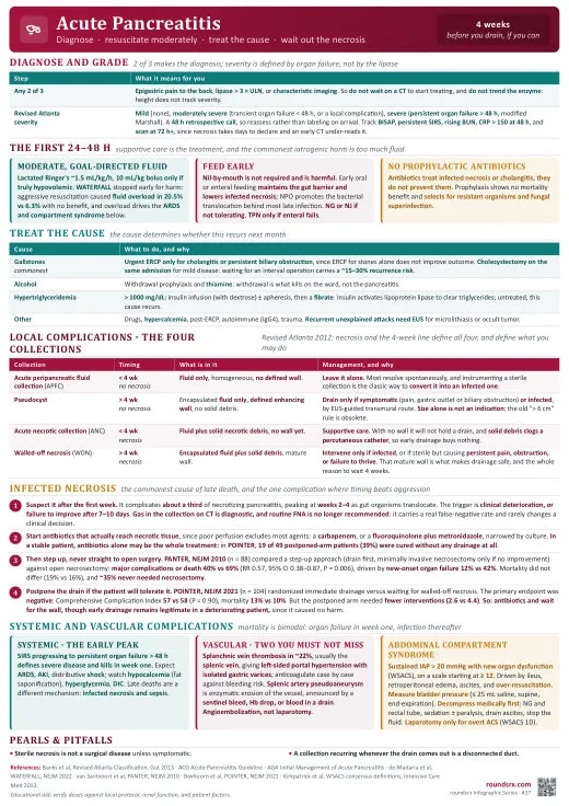
Text version
- Keeping patient NPO (early feeding is safe + beneficial)
- Prophylactic antibiotics (no benefit, promotes resistance)
- Missing gallstone as cause (US + LFTs in all patients)
- Not doing cholecystectomy this admission for gallstone pancreatitis