Radiology Quick Reference
Systematic CXR interpretation, common CT findings, and when to order what. You don't need to be a radiologist -you need to not miss the dangerous stuff.
✅ Is This a Good Film? (RIPE)
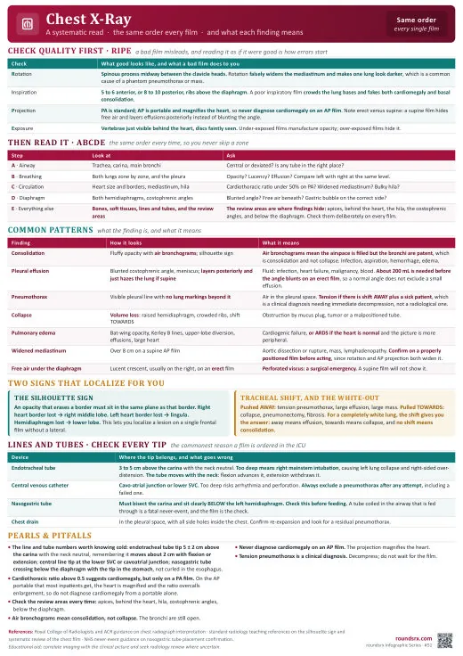
Check adequacy before you interpret. A technically poor film fakes pathology, an underpenetrated or shallow-inspiration film mimics edema and cardiomegaly, a rotated film fakes a widened mediastinum. If you read findings off a bad film, you act on artifact. Run RIPE first, then ABCDE.
| Check | What "good" looks like | If it's off (and how it fools you) |
|---|---|---|
| R -Rotation | The spinous processes sit exactly midway between the medial (sternal) ends of both clavicles. Equal distance = no rotation. Which way: the spinous-to-clavicle gap is wider on the side the patient is rotated toward (it drifts toward the clavicle on the side turned toward the film). | If the spinous process is closer to one clavicle, the patient is rotated. Rotation falsely widens the mediastinum (mimics dissection/mass), makes one lung look denser and the other more lucent (mimics effusion, mass, or pneumothorax), and distorts the heart and hilar contours. Do not call mediastinal width or asymmetric lucency on a rotated film. |
| I -Inspiration | Count 9–10 posterior ribs (or 5–6 anterior ribs) above the diaphragm at the midclavicular line. The diaphragm should sit at about the 10th posterior rib. | Shallow / expiratory film: the bases crowd and look white (mimics edema, basal atelectasis, or infiltrate), the heart is squeezed transversely and looks falsely enlarged, and vascular markings look exaggerated. Hyperexpansion (> 11 ribs, flat diaphragms) points to COPD / air trapping. |
| P -Projection & position | PA, upright, with the side marker present and the scapulae rotated off the lung fields (hands-on-hips technique). Projection = the direction the beam travels (PA, back-to-front; AP, front-to-back). Position = how the patient is placed (upright vs supine vs lateral decubitus). | AP magnifies the heart, so cardiomegaly cannot be called (see box below). Supine loses the air-fluid levels and gravity layering you need to size an effusion or spot a pneumothorax. Lordotic distorts the heart and apices (see box below). On AP/portable the scapulae usually overlie the upper lungs. |
| E -Exposure (penetration) | You should just barely see the lower thoracic vertebrae and disc spaces through the heart, and trace the left hemidiaphragm behind the heart all the way to the spine. Bronchovascular markings visible to within ~2 cm of the pleura. | Underpenetrated (too white): lungs look hazy, mimicking infiltrate or edema; vessels look exaggerated. Overpenetrated (too dark): subtle infiltrates, lines, a small pneumothorax, or a nodule disappear because the markings are burned out. |
Why AP is no good for the heart. On a PA film the beam travels back-to-front and the detector sits against the anterior chest. The heart lies anteriorly, so it is close to the detector and barely magnified, giving a true size (this is why CTR > 0.5 = cardiomegaly only on PA). On an AP film (usually portable, supine, in a sick patient) the detector is behind the patient, so the heart sits far from it and the diverging beam magnifies it by ~15–20%: a normal heart looks big. Supine positioning also raises the diaphragms (lower lung volumes), redistributes flow to the upper lobes (a "cephalization" that is not real failure), and widens the mediastinum. Never diagnose cardiomegaly or mediastinal widening on an AP/portable film; read it as "AP, limited" and correlate.
Lordotic films. Named for lordosis, the spine's normal inward curve; the patient arches back so the beam angles up through the chest. The beam strikes the chest angled upward (patient leaning back, or beam angled cephalad). How to spot it: the medial ends of the clavicles ride high, projected above the lung apices and pointing upward; the anterior ribs look horizontal while the posterior ribs angle steeply; the heart looks foreshortened, rounded, and falsely enlarged; the diaphragm may look flat. How it fools you: the foreshortened heart mimics cardiomegaly, and the apices and RML/lingula are distorted, so you can over-call or miss a consolidation. The useful version: a deliberate apical lordotic view throws the clavicles off the apices to look for apical disease (TB, Pancoast tumor).
🩻 Systematic CXR -ABCDE
| Step | What to Check | Key Findings |
|---|---|---|
| A -Airway | Trachea (midline?), carina, mainstem bronchi | Tracheal deviation: TOWARD collapse/atelectasis, AWAY from tension PTX/large effusion. ETT tip: 2–4 cm above carina. |
| B -Bones & soft tissue | Ribs (fractures?), clavicles, spine, subcutaneous emphysema | Rib fractures → PTX? Flail chest (≥3 consecutive ribs, ≥2 places)? Lytic lesions (mets)? SubQ air → esophageal rupture, necrotizing infection. |
| C -Cardiac | Heart size, silhouette, mediastinum width | CTR > 0.5 = cardiomegaly (on PA film). Boot-shaped → RVH. Water-bottle → pericardial effusion. Widened mediastinum > 8 cm → aortic dissection, aortic aneurysm, lymphoma. |
| D -Diaphragm | Costophrenic angles (blunted?), free air under diaphragm, elevated hemidiaphragm | Blunted CP angles: ≥ 200 mL effusion (lateral decubitus more sensitive). Free air under diaphragm → perforated viscus (surgical emergency). Elevated hemidiaphragm → phrenic nerve palsy, hepatomegaly, subpulmonic effusion. |
| E -Everything else (lungs) | Lung fields: compare L vs R, upper vs lower. Infiltrates, masses, pneumothorax, lines/tubes | White-out: large effusion (meniscus), complete atelectasis (shifted structures toward), massive PNA. PTX: visceral pleural line + absent lung markings. Kerley B lines: pulmonary edema. Check all lines: ETT, central line, NG tube, chest tube, pacer wires. |
Also confirm before reading: right patient, right date, and prior films available for comparison. A single film is a snapshot; the old film tells you what is new.
🔎 Named CXR Signs -What You Actually See
How to use this: the ABCDE tab is how you walk the film; this tab is the lookup for when a pattern jumps out at you. Each row tells you what the sign looks like, then what it means and why, so the finding actually changes your management.
Volume loss / collapse (atelectasis)
| Sign | What you actually see | Pathology & why |
|---|---|---|
| Rib crowding | Rib interspaces on one side visibly narrower than the other; ribs look bunched together | Volume loss. The collapsed lung pulls the chest wall inward, narrowing the gaps. |
| Mediastinal shift toward | Trachea and heart border pulled to the same side as the white opacity | Collapse. Lost volume creates negative space that drags mobile midline structures in. (Shift away = something is pushing: see next table.) |
| Elevated hemidiaphragm | One dome sits higher than expected (normally the right is ~1–2 cm above the left, so look for more than that) | Lower-lobe volume loss (also phrenic palsy, subpulmonic effusion, hepatomegaly). Diaphragm rises to fill the vacated space. |
| Displaced fissure | The thin white line of a fissure pulled out of its normal position toward the opacity | Lobar collapse. The fissure migrates with the shrinking lobe; the most direct sign of which lobe collapsed. |
Pushing pathology (volume added)
| Sign | What you actually see | Pathology & why |
|---|---|---|
| Mediastinal shift away | Trachea and heart pushed to the opposite side of the white-out | Large effusion or mass. Volume is being added and shoves the mediastinum off midline. Pairs with the meniscus sign for effusion. |
| Deep sulcus sign | On a supine film, one costophrenic angle is abnormally deep and very black, with a sharp lateral gutter | Pneumothorax. In a supine patient, air rises anterobasally (the least dependent spot) and deepens the sulcus, where a classic apical pleural line may be absent. |
| Visible pleural line + absent markings | A thin white line parallel to the chest wall with pure black (no vascular markings) beyond it | Pneumothorax. The line is the visceral pleura; the black void is free pleural air with no lung in it. |
| Trachea away + flat/low diaphragm | Everything pushed off midline, hemidiaphragm flattened or pushed down | Tension pneumothorax. Air under pressure pushes the mediastinum and kinks venous return. This is a clinical diagnosis; decompress before you wait on the film. |
Air-space, interstitial & pleural
| Sign | What you actually see | Pathology & why |
|---|---|---|
| Air bronchogram | Dark, branching tube-like lucencies running through a white patch of lung | Alveolar filling (pneumonia, pulmonary edema, ARDS). Alveoli fill with fluid or pus but the bronchi stay air-filled, so they show up dark against the white. |
| Silhouette sign | A border that should be a crisp line (heart edge or diaphragm) is blurred or erased | Localizes the disease. Two same-density structures in contact lose the line between them: lost right heart border = RML, lost left heart border = lingula, lost diaphragm outline = lower lobe. |
| Kerley B lines | Short (1–2 cm), thin horizontal lines at the periphery, perpendicular to and touching the pleural surface (usually at the bases) | Interstitial edema or lymphangitic spread. Fluid or cells thicken the interlobular septa enough to be seen. |
| Bat-wing / butterfly | Symmetric fluffy white haze fanning out from both hila, sparing the periphery | Cardiogenic pulmonary edema. Fluid collects centrally first before reaching the outer lung. |
| Meniscus sign | A white fluid density whose upper edge curves upward (concave-up) along the lateral chest wall | Pleural effusion. Free fluid tracks up the dependent space; surface tension gives the upward curve. Blunted costophrenic angle is the earliest version (~200 mL on an upright film). |
Classic spot-diagnoses
| Sign | What you actually see | Pathology & why |
|---|---|---|
| Free air under diaphragm | A thin black crescent of air between the white diaphragm and the liver, on an upright film | Perforated viscus (surgical emergency). Free intraperitoneal air rises under the dome. Needs an upright or left-lateral-decubitus film to be seen. |
| Continuous diaphragm sign | The full diaphragm line is visible across the midline, including the central part normally hidden by the heart | Pneumomediastinum. Air outlines the central diaphragm from above, completing the line. |
| Westermark sign | A focal region of lung that looks darker (fewer vessels) than the rest | Pulmonary embolism. Clot causes oligemia distal to it. Insensitive but a classic boards answer. |
| Hampton hump | A wedge-shaped white density at the lung periphery, base on the pleura, apex pointing toward the hilum | PE with infarction. The wedge is the infarcted lung territory. |
| Boot-shaped heart | Cardiac apex lifted up off the diaphragm with a concave upper-left border, shaped like a boot | Tetralogy of Fallot. RV hypertrophy tilts the apex upward. |
| Water-bottle heart | Globular, symmetrically enlarged cardiac silhouette with a smooth flask shape | Large pericardial effusion. Fluid in the pericardial sac balloons the silhouette; correlate with echo, not CXR alone. |
🔍 Common CT Findings
| Finding | Think | Clinical Action |
|---|---|---|
| CT Head -hyperdense (white) | Acute blood (ICH, SDH, EDH, SAH) | SDH: crescent-shaped, crosses suture lines. EDH: lens-shaped, doesn't cross sutures. SAH: blood in sulci/cisterns → CTA for aneurysm. ICH: check INR, reverse anticoag. |
| CT Head -hypodense (dark) | Ischemic stroke (after 6–12h), chronic SDH, edema | Acute stroke may be normal on CT < 6h → loss of gray-white differentiation is earliest sign. MRI DWI is more sensitive early. |
| CTPA -filling defect in PA | Pulmonary embolism | Saddle PE (at bifurcation) → hemodynamically significant. RV/LV ratio > 1 → RV strain. Check troponin, BNP. |
| CT Chest -ground glass opacities (GGO) | PCP pneumonia (HIV), viral PNA (COVID, influenza), pulmonary edema, DAH, early ILD, drug toxicity | Bilateral diffuse GGO + HIV → check CD4, start TMP-SMX for PCP. Bilateral + ICU → ARDS. Peripheral/basal → UIP/IPF pattern. |
| CT Chest -tree-in-bud pattern | TB, atypical mycobacteria, aspiration | Small airway disease. Centrilobular nodules + tree-in-bud = active infectious bronchiolitis. Isolate for TB. Get sputum AFB × 3. |
| CT Abdomen -free air | Perforated viscus | Surgical emergency. Most common: perforated duodenal ulcer, perforated diverticulitis, perforated appendix. Get surgical consult immediately. |
| CT Abdomen -portal venous gas | Mesenteric ischemia, bowel necrosis | Ominous sign. Also pneumatosis intestinalis (air in bowel wall). Lactate elevated. Surgical emergency. CT angiography for mesenteric vessels. |
| CT Abdomen -dilated bowel | SBO vs ileus | SBO: transition point (dilated → decompressed), small bowel > 3 cm. Ileus: diffusely dilated, no transition point. SBO → surgical consult. Toxic megacolon (C. diff): colon > 6 cm. 3-6-9 rule for upper limits: small bowel 3, colon 6, cecum 9 cm (3-6-9 rule). |
📋 What Imaging to Order
| Clinical Question | Best Study | Notes |
|---|---|---|
| Chest pain -ACS ruled out, PE suspected | CTPA | Wells score first. If low probability → D-dimer. If D-dimer positive or high probability → CTPA. V/Q scan if contrast allergy or CKD. |
| Stroke symptoms (< 24h) | CT Head without contrast (rule out hemorrhage) → CTA head/neck (LVO) → MRI DWI (confirm ischemic) | CT to rule out bleed before tPA. CTA for large vessel occlusion (thrombectomy candidate). MRI DWI most sensitive for acute ischemia. |
| Abdominal pain | CT abdomen/pelvis with IV contrast | Most versatile. RLQ pain in young female → consider US first (ovarian pathology, avoid radiation). RUQ pain → RUQ US first (gallstones, cholecystitis). |
| GI bleed -upper vs lower | EGD first. CT angiography if massive/unstable. | CTA abdomen/pelvis if active bleeding (extravasation). Tagged RBC scan if slow intermittent bleed. Colonoscopy within 24h for lower GIB. |
| Renal colic / stones | CT abdomen/pelvis WITHOUT contrast | Non-contrast CT is gold standard for stones (contrast obscures them). US is first-line in pregnancy. |
| Biliary disease | RUQ ultrasound | First-line for gallstones, cholecystitis. MRCP for common bile duct stones if US equivocal. HIDA scan for acalculous cholecystitis (EF < 35%). |
| DVT suspected | Compression ultrasound | Sensitivity > 95% for proximal DVT. If negative but high clinical suspicion → repeat in 5–7 days or whole-leg US. |
| Aortic dissection | CTA chest/abdomen/pelvis | Gold standard. TEE is alternative (especially if too unstable for CT). D-dimer < 500 has high NPV for dissection. |
| Pleural effusion workup | CXR → US-guided thoracentesis | Lateral decubitus CXR to confirm free-flowing. Bedside US for marking. CT chest with contrast if concern for malignancy, empyema, or loculated. |
| Pulmonary nodule found | Follow Fleischner criteria | Size, morphology (solid vs GGO vs part-solid), risk factors determine follow-up interval. Part-solid nodules have highest malignancy risk. PET-CT for solid nodules ≥ 8 mm. |
Contrast contraindications: eGFR < 30 (risk of CIN -but don't withhold if life-threatening indication like PE, dissection, stroke), contrast allergy (premedicate: prednisone 50 mg at 13h, 7h, 1h before + diphenhydramine 50 mg 1h before). Metformin: hold day of contrast, resume 48h later if Cr stable.