Amyloidosis
Amyloidosis is a group of diseases caused by extracellular deposition of misfolded proteins (amyloid fibrils) in tissues, leading to organ dysfunction. The protein type determines the disease subtype and treatment.
- AL Amyloidosis (Light Chain) -from plasma cell dyscrasia. Most common systemic amyloidosis. Light chains (usually lambda) misfold and deposit. Related to multiple myeloma but most patients do NOT have overt myeloma.
- AA Amyloidosis -from serum amyloid A (SAA) protein, produced in chronic inflammation. Causes: RA, Crohn disease, familial Mediterranean fever (FMF), chronic infections. Predominantly affects kidneys.
- ATTR Amyloidosis (Transthyretin) -hereditary (mutant TTR) or wild-type (senile cardiac amyloidosis). Increasingly recognized as a cause of HFpEF in elderly patients.
- Heart -restrictive cardiomyopathy. Low voltage on ECG despite thick walls on echo (classic discordance). Diastolic dysfunction. Elevated troponin and BNP.
- Kidney -nephrotic syndrome (proteinuria, edema, hypoalbuminemia)
- Liver -hepatomegaly, elevated ALP
- Nerves -peripheral neuropathy (pain, numbness), autonomic neuropathy (orthostatic hypotension)
- GI -malabsorption, weight loss, GI bleeding
- Tongue -macroglossia (pathognomonic for AL amyloidosis)
- Treat the underlying plasma cell clone -goal is to eliminate the source of amyloidogenic light chains.
- First-line: Daratumumab + CyBorD (cyclophosphamide, bortezomib, dexamethasone). ANDROMEDA, NEJM 2021 -showed significantly higher hematologic complete response rate.
- Autologous stem cell transplant (SCT) -for eligible patients (typically younger, no significant cardiac involvement). Best long-term outcomes.
- Cardiac AL prognosis: Very poor -median survival 6 months untreated. With modern treatment, outcomes improving but cardiac involvement remains the dominant prognostic factor.
Treat the underlying inflammatory disease. Control RA, Crohn, FMF (colchicine for FMF). Reducing SAA levels can halt progression and even allow regression of amyloid deposits.
- Heart failure: Diuretics -use cautiously, these patients are preload-dependent (restrictive physiology). Small changes in volume cause dramatic hemodynamic effects. Avoid digoxin (binds amyloid fibrils, causing toxicity at therapeutic levels).
- Orthostatic hypotension: Midodrine 5-10 mg TID, compression stockings, increased salt intake.
- Nephrotic syndrome: ACEi/ARB for proteinuria. Diuretics for edema.
- Tissue biopsy with Congo red stain -apple-green birefringence under polarized light is diagnostic of amyloid.
- Fat pad aspirate -least invasive biopsy site. Sensitivity ~70-80% for AL. If negative but suspicion high, biopsy the affected organ.
- Bone marrow biopsy -to assess plasma cell clone in AL amyloidosis.
- SPEP / UPEP / serum free light chains -for AL amyloidosis. Free light chain ratio is abnormal in >95% of AL patients.
- SAA levels -for AA amyloidosis. Elevated in chronic inflammatory states.
- Mass spectrometry -gold standard for amyloid subtyping (laser microdissection + mass spec on biopsy tissue).
- Troponin and BNP/NT-proBNP -markedly elevated in cardiac amyloidosis. Used for staging (Mayo staging system).
- ECG -low voltage (limb leads <5mm) + pseudo-infarct pattern (Q waves without MI) are classic.
- Echocardiogram -thick walls + diastolic dysfunction. "Sparkling" or "granular" myocardium (sometimes described on older machines). Small LV cavity, biatrial enlargement.
- Cardiac MRI -characteristic diffuse subendocardial or transmural late gadolinium enhancement pattern. Global T1 elevation.
- Technetium pyrophosphate (PYP) scan -highly sensitive and specific for ATTR cardiac amyloidosis. Grade 2-3 uptake = diagnostic without biopsy.
| Drug | Type | Dose | Notes |
|---|---|---|---|
| Daratumumab (Darzalex) AL 1ST LINE | AL | 16 mg/kg IV weekly x 8, then q2 weeks, then monthly | Anti-CD38 monoclonal antibody. Combined with CyBorD. ANDROMEDA, NEJM 2021 |
| Bortezomib (Velcade) | AL | 1.3 mg/m2 SQ weekly (part of CyBorD) | Proteasome inhibitor. Targets plasma cells. Peripheral neuropathy is dose-limiting toxicity. |
| Cyclophosphamide | AL | 300 mg/m2 PO weekly (part of CyBorD) | Alkylating agent. Dose adjust for renal function. |
| Dexamethasone | AL | 20-40 mg PO weekly (part of CyBorD) | Steroid component. Monitor glucose, infections. |
| Tafamidis (Vyndamax) | ATTR | 80 mg PO daily | TTR stabilizer for ATTR cardiac amyloidosis. Reduced mortality and CV hospitalization. ATTR-ACT, NEJM 2018 |
| Midodrine | Supportive | 5-10 mg PO TID | For orthostatic hypotension from autonomic neuropathy. Hold if supine BP >180. |
Patient: 64M with progressive dyspnea, bilateral LE edema, and orthostatic hypotension. Echo: thick walls (IVS 16 mm), small LV cavity, diastolic dysfunction, EF 50%. ECG: low voltage + pseudo-infarct pattern. NT-proBNP 9200, troponin 0.18. Macroglossia on exam.
Key findings: Classic cardiac AL amyloidosis: thick walls + low voltage on ECG = pathognomonic discordance (hypertrophy causes high voltage, but amyloid infiltration causes low voltage). Macroglossia is virtually diagnostic for AL type.
Management:
- Fat pad aspirate or organ biopsy → Congo red stain (apple-green birefringence under polarized light)
- Mass spectrometry typing to confirm AL (vs ATTR), critical for treatment selection
- Dara-CyBorD: daratumumab + bortezomib + cyclophosphamide + dexamethasone (first-line per ANDROMEDA) ANDROMEDA, 2021
- Diuretics for volume overload, use cautiously (preload-dependent restrictive physiology). AVOID digoxin (binds to amyloid fibrils → toxicity at therapeutic levels)
- Midodrine for orthostatic hypotension (autonomic neuropathy from amyloid infiltration)
Teaching point: The ECG-echo discordance (low voltage + thick walls) is the signature finding of cardiac amyloidosis. HCM and hypertensive heart disease cause thick walls WITH high voltage. If the walls are thick but voltage is low → think amyloid.
Patient: 82M with HFpEF refractory to standard therapy. History of bilateral carpal tunnel surgery, lumbar spinal stenosis. Echo: concentric LVH (IVS 15 mm), grade III diastolic dysfunction. Technetium pyrophosphate scan: grade 3 uptake. SPEP/sFLC normal.
Key findings: Wild-type ATTR (ATTRwt), transthyretin amyloidosis from age-related misfolding. No plasma cell dyscrasia (SPEP/sFLC normal). Tc-PYP scan grade 2-3 = diagnostic for ATTR without biopsy (if SPEP/sFLC negative). Bilateral carpal tunnel + spinal stenosis are early manifestations.
Management:
- Tafamidis 80 mg daily, TTR stabilizer, reduces mortality and HF hospitalization ATTR-ACT, 2018
- No chemotherapy needed (ATTR is not a plasma cell disorder, no clonal cells to target)
- Diuretics for congestion, careful volume management
- Genetic testing: rule out hereditary ATTR (TTR gene mutations, affects family screening and transplant eligibility)
- Avoid ACEi/ARB aggressively, often poorly tolerated due to restrictive physiology + autonomic dysfunction
Teaching point: ATTR is now recognized as a common cause of HFpEF in elderly men, prevalence may be 10-15% of HFpEF patients > 80. Bilateral carpal tunnel + HFpEF + thick walls should trigger Tc-PYP scan. Tafamidis is disease-modifying.
Patient: 58F with progressive LE edema, foamy urine. Albumin 1.8, 24h urine protein 8.2 g/day, Cr 1.6. sFLC: lambda 420 mg/L (elevated), ratio abnormal. Renal biopsy: Congo red positive in glomeruli and vessels.
Key findings: AL amyloidosis presenting as nephrotic syndrome (renal is the most common organ involved in AL). Lambda light chain predominance. Must assess cardiac involvement, asymptomatic cardiac amyloid can coexist.
Management:
- Cardiac biomarkers: NT-proBNP + troponin (Mayo staging, determines prognosis and treatment intensity)
- Echo + ECG to assess subclinical cardiac involvement
- Dara-CyBorD (standard first-line for AL amyloidosis regardless of organ involvement)
- Aggressive diuresis for nephrotic edema (furosemide + metolazone). ACEi/ARB for proteinuria reduction
- If young + fit + good cardiac function: consider autologous stem cell transplant after induction (high CR rates in selected patients)
Teaching point: In any patient with unexplained nephrotic syndrome + monoclonal protein, think AL amyloidosis. The combination of nephrotic-range proteinuria + abnormal sFLC ratio is the classic presentation. Always check cardiac biomarkers, occult cardiac involvement determines prognosis.
Mr. Franklin is a 62-year-old man presenting with 6 months of progressive dyspnea on exertion, lower extremity edema, and 15-lb weight loss. He also reports numbness/tingling in both feet and lightheadedness when standing. Exam: macroglossia, periorbital purpura, elevated JVP, bilateral pitting edema. Labs: troponin 0.15 (elevated), NT-proBNP 8,400 (markedly elevated), creatinine 1.8, albumin 2.1, 24h urine protein 5.2 g/day. ECG: low voltage + pseudo-infarct pattern. Echo: thick walls (IVS 16mm) + diastolic dysfunction + small LV cavity. Free kappa/lambda ratio markedly abnormal (lambda predominant). Fat pad biopsy: Congo red positive, apple-green birefringence under polarized light.
- Serum free light chains -q1-3 months during treatment. Hematologic response: normalization of free light chain ratio.
- NT-proBNP and troponin -cardiac biomarkers used for staging and monitoring organ response. Reduction indicates cardiac response.
- 24h urine protein -for renal involvement. Renal response: >50% reduction in proteinuria.
- Echocardiogram -q6-12 months. Monitor wall thickness, diastolic function, strain patterns.
- ALP -for hepatic involvement. Hepatic response: >50% decrease in ALP.
- Orthostatic vitals -for autonomic neuropathy monitoring.
- CBC, BMP -monitor for treatment toxicity (myelosuppression from chemo, renal function).
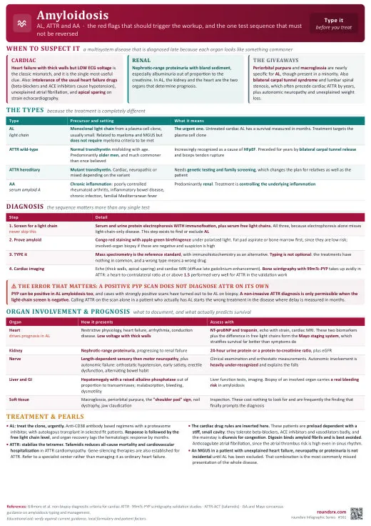
Text version
- AL (light chain): from a plasma cell dyscrasia. The most common systemic amyloidosis. Light chains, usually lambda, misfold and deposit. Related to multiple myeloma, but most patients do not have overt myeloma, so a normal marrow does not exclude it. Treatment targets the clone.
- ATTR (transthyretin), hereditary: mutant TTR. Genetic testing matters here because it changes family screening and transplant eligibility, not just the label.
- ATTR, wild-type (ATTRwt, senile cardiac amyloidosis): age-related TTR misfolding with no plasma cell clone. Increasingly recognized as a cause of HFpEF in elderly patients; prevalence may be 10–15% of HFpEF patients > 80. Bilateral carpal tunnel surgery and lumbar spinal stenosis are early manifestations that precede the heart failure by years.
- AA: from serum amyloid A (SAA) produced in chronic inflammation. Causes: RA, Crohn disease, familial Mediterranean fever, chronic infections. Predominantly affects the kidneys. Treatment is control of the inflammatory driver, not chemotherapy.
- Do not blur AL and ATTR. AL gets chemotherapy; ATTR has no clone to target and gets a TTR stabilizer. Getting this backwards means either useless chemotherapy or an untreated fatal plasma cell disease.
- Heart, restrictive cardiomyopathy: low voltage on ECG despite thick walls on echo, the classic voltage-mass discordance. In HCM and hypertensive heart disease, thick walls produce high voltage because the thickness is myocyte hypertrophy. In amyloid, the thickness is electrically inert protein infiltrating between myocytes, which insulates the myocardium.
- Kidney: nephrotic syndrome, proteinuria, edema, hypoalbuminemia. Unexplained nephrotic syndrome plus a monoclonal protein is AL until proven otherwise.
- Tongue, macroglossia: pathognomonic for AL, present in roughly 10–15% of AL patients and does not occur in AA or ATTR. Look for tooth indentations on the lateral tongue edges; it can also cause dysphagia and obstructive sleep apnea.
- Nerves: peripheral neuropathy (pain, numbness) and autonomic neuropathy (orthostatic hypotension). The orthostasis is not just deconditioning.
- Liver: hepatomegaly with elevated alkaline phosphatase.
- GI: malabsorption, weight loss, GI bleeding.
- Serum free light chains q1–3 months during treatment. Hematologic response means normalization of the free light chain ratio, and it moves before organ function does.
- NT-proBNP and troponin for staging and organ response; a reduction indicates cardiac response, the response that matters most for survival.
- 24h urine protein for renal involvement. Renal response is defined as a > 50% reduction in proteinuria.
- ALP for hepatic involvement; hepatic response is a > 50% decrease.
- Echocardiogram q6–12 months to track wall thickness, diastolic function, and strain patterns.
- Orthostatic vitals to follow autonomic neuropathy and titrate midodrine.
- CBC and BMP for treatment toxicity: myelosuppression from chemotherapy and renal function.
- Digoxin in cardiac amyloidosis. It binds amyloid fibrils and accumulates in tissue, producing toxicity and potentially fatal arrhythmias at "therapeutic" serum levels. Avoid it entirely.
- Aggressive diuresis. Restrictive physiology makes these patients preload-dependent, so small volume changes cause dramatic hemodynamic effects. Diurese, but cautiously.
- Reading thick walls as HCM or hypertensive heart disease. Thick walls with low voltage is amyloid; thick walls with high voltage is hypertrophy.
- Skipping the typing step. Congo red only says "amyloid". Committing an ATTR patient to chemotherapy, or an AL patient to tafamidis alone, follows directly from that shortcut.
- Calling a PYP scan diagnostic while the clone workup is still open. Grade 2–3 uptake confirms ATTR without biopsy only when SPEP and sFLC are negative, since a coexisting monoclonal protein reopens the AL question.
- Assuming no myeloma means no AL. Most AL patients do not have overt myeloma.
- Assuming renal-only disease. Asymptomatic cardiac amyloid can coexist and determines prognosis, so check cardiac biomarkers even when the presentation is purely nephrotic.
- Pushing ACEi/ARB hard in ATTR cardiac amyloid; they are often poorly tolerated because of restrictive physiology plus autonomic dysfunction.
- AL: treat the underlying plasma cell clone to eliminate the source of amyloidogenic light chains. First-line is Dara-CyBorD (daratumumab plus cyclophosphamide, bortezomib, dexamethasone), regardless of which organ is involved.
- AL, transplant-eligible: autologous stem cell transplant for younger, fit patients without significant cardiac involvement, which gives the best long-term outcomes; consider after induction.
- Cardiac AL is the prognostic driver: median survival 6 months untreated. Elevated troponin plus NT-proBNP means advanced cardiac involvement by Mayo staging and should accelerate, not delay, referral.
- ATTR: tafamidis 80 mg daily. No chemotherapy, there is no clonal cell population to target.
- AA: treat the underlying inflammatory disease (RA, Crohn, FMF with colchicine). Lowering SAA can halt progression and even allow regression of deposits, which is why disease control is the therapy.
- New complete heart block, syncope, or refractory heart failure: cardiology involvement; restrictive physiology limits standard HF therapy.
- Bilateral carpal tunnel syndrome plus HFpEF plus thick walls in an older patient: order the Tc-PYP scan rather than continuing to titrate diuretics.
- ANDROMEDA, NEJM 2021: adding daratumumab to CyBorD in newly diagnosed AL doubled the hematologic complete response (53% vs 18%) and improved cardiac and renal organ response. Dara-CyBorD is now the first-line standard.
- ATTR-ACT, NEJM 2018: tafamidis reduced all-cause mortality and cardiovascular hospitalization in ATTR cardiomyopathy, making it genuinely disease-modifying rather than supportive.
- Mayo staging using troponin and NT-proBNP is what converts "cardiac involvement" into a prognosis and a treatment intensity, so send both at diagnosis.