Anemia Workup
| MCV | Category | Differential | Key Labs |
|---|---|---|---|
| < 80 | Microcytic | Iron deficiency (#1 -GI blood loss in men/postmenopausal women until proven otherwise), thalassemia (Mentzer index: MCV/RBC < 13 → thalassemia, > 13 → iron def), anemia of chronic disease (some), sideroblastic, lead poisoning | Iron studies, ferritin, TIBC, reticulocyte count, Hgb electrophoresis if thalassemia suspected |
| 80–100 | Normocytic | Anemia of chronic disease (#1), acute blood loss, CKD (↓ EPO), mixed deficiency (iron + B12), hemolysis (check hemolysis labs), bone marrow failure (aplastic, MDS, infiltration) | Reticulocyte count (↑ = destruction/loss, ↓ = underproduction), BMP (CKD), hemolysis labs (LDH, haptoglobin, indirect bili, smear) |
| > 100 | Macrocytic | B12 deficiency (neurologic symptoms -subacute combined degeneration; ask about nitrous oxide abuse, where the B12 level is usually normal and the CBC is usually normal too), folate deficiency, alcohol/liver disease, hypothyroidism, MDS, medications (methotrexate, hydroxyurea, azathioprine, AZT), reticulocytosis (reticulocytes are large → MCV ↑) | B12, folate, reticulocyte count, TSH, peripheral smear (hypersegmented neutrophils → megaloblastic), methylmalonic acid (↑ in B12 def, normal in folate def) |
- Reticulocyte count > 2% (or reticulocyte index > 2): bone marrow is responding → the problem is blood loss or destruction (hemolysis)
- Reticulocyte count < 2%: bone marrow is NOT responding → underproduction (iron/B12/folate deficiency, CKD, bone marrow failure, anemia of chronic disease)
- Reticulocyte Production Index (RPI): (retic % × Hgb/15) / maturation factor. RPI > 2 = appropriate response.
| Lab | Iron Deficiency | Anemia of Chronic Disease | Both (Mixed) |
|---|---|---|---|
| Serum iron | ↓ | ↓ | ↓ |
| Ferritin | ↓↓ (< 30) | ↑ or normal (acute phase reactant) | Low-normal (30–100) |
| TIBC | ↑ (body wants more iron) | ↓ (body not trying to absorb more) | Variable |
| Transferrin saturation (TSAT) | ↓ (< 20%) | ↓ (15–20%) | ↓ |
Why it complements ferritin: TSAT reflects the iron actually available right now for erythropoiesis (the "circulating" iron pool), while ferritin reflects storage iron. TSAT is less affected by inflammation, so it's the more reliable marker when ferritin is unreliable -hospitalized inflamed patients, HF, CKD, chronic infection, malignancy. A patient with ferritin 80 + TSAT 12% is iron-deficient even though the ferritin looks "OK."
Pre-test pearls: hold oral iron for 24-48 h before drawing labs (recent iron pill falsely elevates TSAT). TSAT has a diurnal rhythm -higher in the morning -so a fasting AM draw is most reliable when the result is borderline.
| Route | Agent | Notes |
|---|---|---|
| Oral | Ferrous sulfate 325 mg PO daily–TID | Take on empty stomach with vitamin C (enhances absorption). GI side effects are dose-limiting. Every-other-day dosing may improve absorption and tolerability. Takes 3–6 months to replete stores. |
| IV (preferred if) | Iron sucrose (Venofer) 200 mg IV × 5 doses or ferric carboxymaltose (Injectafer) 750 mg IV × 2 | Preferred if: oral intolerant, CKD/dialysis, IBD, ongoing blood loss exceeding oral repletion, Hgb < 7 with symptoms, pre-surgery. Injectafer: can give 750 mg in one sitting (fewer visits). |
| Test | What You Are Looking For | Why It Matters |
|---|---|---|
| CBC with reticulocyte count FIRST | The MCV sorts the cause by size (microcytic, normocytic, macrocytic) and the retic sorts it by mechanism. | Correct the retic for the degree of anemia, since a raw percentage of a small red cell mass overstates the response. An untreated iron deficiency with a "normal" retic is a marrow that is failing to respond, not one that is coping. |
| Peripheral smear DO NOT SKIP | Schistocytes, spherocytes, bite cells, target cells, hypersegmented neutrophils, teardrops, blasts, rouleaux. | ⚠ Schistocytes change the night. They point to a microangiopathic process, and TTP is a hematologic emergency where the delay to plasma exchange is what kills. Hypersegmented neutrophils flag B12 or folate deficiency before the MCV rises, and blasts mean the diagnosis is leukemia, not anemia. No analyzer index substitutes for looking. |
| Iron studies | Ferritin < 30 ng/mL is diagnostic of iron deficiency (sensitivity 92%, specificity 98%). Transferrin saturation < 20% supports it. | ⚠ Ferritin is an acute phase reactant, so inflammation can lift it into the normal range in a genuinely iron-deficient patient. In chronic inflammation, raise the cutoff toward 100 ng/mL and lean on transferrin saturation instead. This is the commonest way iron deficiency gets missed on a normal-looking panel. |
| B12 and folate | B12 below roughly 200 pg/mL. Check methylmalonic acid and homocysteine when the level is borderline, and always if nitrous oxide is in the history, where B12 is only 22% sensitive but homocysteine and MMA are over 90% (why). | The borderline zone is common and the metabolites settle it, because MMA rises in true B12 deficiency and stays normal in folate deficiency. It matters because giving folate alone to a B12-deficient patient corrects the anemia while the neurologic damage progresses, and that damage can be permanent. |
| Hemolysis panel | LDH high, haptoglobin low, indirect bilirubin high, retic high. All four move together. | Haptoglobin is the most useful single member: it is consumed by free hemoglobin, so a low haptoglobin with a high LDH is strong evidence of hemolysis. Interpret them as a set, since LDH alone rises in almost anything. |
| Direct antiglobulin test (Coombs) | Antibody or complement on the red cell surface. | Splits confirmed hemolysis into immune and non-immune, which is the branch point for treatment. Positive points to autoimmune hemolysis and steroids; negative redirects you to microangiopathy, a mechanical valve, G6PD or a membrane defect. |
| Renal function and EPO | Creatinine, eGFR, and an erythropoietin level where the picture is unclear. | Anemia of chronic kidney disease is an erythropoietin deficiency, and it is common enough to explain many normocytic anemias without further hunting. Correct iron first, since an ESA will not work in an iron-deficient patient. |
| Bone marrow biopsy | Reserved for when the non-invasive workup does not explain it. | Indicated for pancytopenia, circulating blasts, suspected myelodysplasia or infiltration. It is the answer to an unexplained hypoproliferative anemia, not a first-line test. |
- Iron deficiency: FIND SOURCE + IV iron (ferric carboxymaltose 750mg×2) or PO. Transfuse: Hgb<7 in most patients TRICC, Hébert 1999
- B12: IM cyanocobalamin 1000mcg daily×7→weekly×4→monthly
- Hemolytic: autoimmune→steroids±rituximab. TTP→PLEX. Mechanical→treat cause.
- CKD: iron first, then ESA (Hgb 10-11)
- Transfuse: Hgb<7 most, <8 ACS TRISS, Holst 2014 TITRe2, Murphy 2015
Patient: 34-year-old premenopausal woman with heavy menstrual periods presents with fatigue and pica (ice craving).
Key findings: Hgb 7.6, MCV 65, ferritin 4, TIBC 520, TfSat 4%, reticulocyte index 0.5%. Smear: microcytic hypochromic cells, pencil cells.
Management:
- IV iron (ferric carboxymaltose 750 mg x 2) due to severity
- Gynecology referral for menorrhagia management
- GI workup deferred given clear gynecologic source and young age
Teaching point: Pica (pagophagia) is specific for iron deficiency. In premenopausal women, menorrhagia is the most common cause. IRON-MIDE, 2020
Patient: 42-year-old woman with SLE presents with jaundice, dark urine, and worsening fatigue. Splenomegaly on exam.
Key findings: Hgb 6.2, MCV 102 (elevated from reticulocytosis), reticulocyte count 12%, LDH 680, haptoglobin undetectable, indirect bilirubin 4.8, DAT positive (IgG + C3).
Management:
- Warm AIHA secondary to SLE: prednisone 1 mg/kg/day
- Transfuse if hemodynamically unstable
- If steroid-refractory: rituximab or splenectomy
Teaching point: Warm AIHA (IgG-mediated) is associated with SLE, CLL, and drugs. MCV may be falsely elevated because reticulocytes are larger than mature RBCs. Haptoglobin is the most sensitive hemolysis marker.
Patient: 68-year-old vegan woman presents with fatigue, glossitis, and bilateral lower extremity paresthesias. Decreased vibration sense, positive Romberg.
Key findings: Hgb 9.2, MCV 124, reticulocyte index 0.6. B12 less than 100. Methylmalonic acid 2400 (markedly elevated). Smear: macro-ovalocytes, hypersegmented neutrophils.
Management:
- IM cyanocobalamin 1000 mcg daily x 7d, then weekly x 4, then monthly lifelong
- Monitor K+ and phosphate (drop during rapid erythropoiesis)
- Neurology follow-up for subacute combined degeneration
Teaching point: B12 deficiency causes subacute combined degeneration (posterior columns + lateral corticospinal tracts). Methylmalonic acid differentiates B12 from folate deficiency. Neurologic damage may be irreversible if treatment is delayed.
| Drug | Dose | Route | Indication |
|---|---|---|---|
| Ferrous sulfate | 325 mg PO daily–TID | PO | Iron deficiency anemia (mild-moderate). Take on empty stomach with vitamin C. Every-other-day dosing may improve absorption. |
| Iron sucrose (Venofer) | 200 mg IV x 5 doses | IV | IV iron preferred if: oral intolerant, CKD/dialysis, IBD, Hgb < 7, ongoing blood loss, pre-surgery. |
| Ferric carboxymaltose (Injectafer) | 750 mg IV x 2 (1 week apart) | IV | Convenient IV iron -can give 750 mg in one sitting (fewer visits). |
| Cyanocobalamin (B12) | 1000 mcg IM daily x 7d → weekly x 4 → monthly | IM | B12 deficiency. High-dose oral (1000–2000 mcg/day) may be adequate if no absorption issues. |
| Folic acid | 1 mg PO daily | PO | Folate deficiency. Always check B12 first -folate alone can mask B12 deficiency (corrects anemia but NOT neuro damage). |
| Epoetin alfa (Procrit) | 50–300 units/kg 3x/week | IV/SQ | Anemia of CKD (after iron repletion). Target Hgb 10–11.5. Never > 13. |
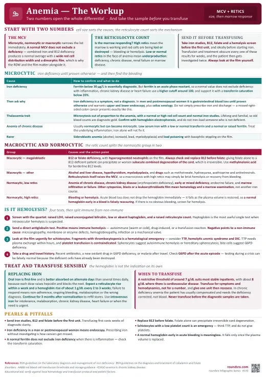
Text version
- Transfusing above Hgb 7 threshold without indication
- Not checking reticulocyte count (production vs destruction)
- Missing B12 deficiency (can cause pancytopenia mimicking leukemia)
- Iron infusion without finding the source of loss