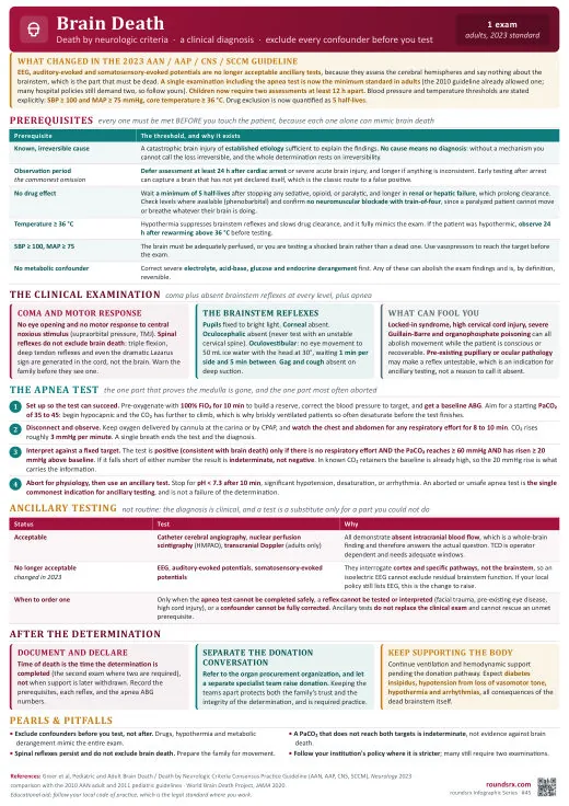
Brain Death Evaluation
| Prerequisite | Details |
|---|---|
| Known cause | Established etiology sufficient to cause brain death (e.g., massive stroke, TBI, anoxic injury) |
| Irreversibility | No possibility of recovery -adequate time for observation |
| No confounders | Core temp ≥ 36°C, SBP ≥ 100, no CNS depressants (check drug levels), no neuromuscular blockade, no severe metabolic derangements |
| Etiology | Frequency | Key Features |
|---|---|---|
| Traumatic brain injury | ~40% | MVA, falls, assaults. Often young patients. CT: diffuse axonal injury, herniation |
| Hemorrhagic stroke | ~25% | Massive ICH, SAH with rebleed. CT: large hematoma with midline shift, herniation |
| Anoxic brain injury | ~20% | Cardiac arrest, drowning, strangulation. CT: diffuse edema, loss of gray-white differentiation |
| Ischemic stroke | ~10% | Large territory (MCA/basilar). CT: massive hemispheric or posterior fossa infarct with edema |
| Other | ~5% | CNS infection, tumor, fulminant hepatic failure with cerebral edema |
- Attending physician with experience in brain death determination (varies by state law and hospital policy)
- Typically: neurologist, neurosurgeon, or intensivist
- The physician performing the evaluation should NOT be part of the transplant team (conflict of interest) Uniform Determination of Death Act, 1981
- Two separate exams recommended (2010 AAN); 2023 guidelines state one exam by one qualified physician is sufficient in adults
| Brainstem Reflex | Test | Expected in Brain Death |
|---|---|---|
| Pupillary | Bright light in each eye | Fixed, dilated (4–9 mm), no response |
| Corneal | Cotton wisp or saline drops | No blink |
| Oculocephalic | Doll's eyes (turn head side to side) | No eye movement (eyes stay midline) |
| Oculovestibular | Cold caloric (50 mL ice water in ear canal) | No eye deviation toward cold ear |
| Gag | Stimulate posterior pharynx | No response |
| Cough | Suction catheter to carina | No cough |
- Pre-oxygenate with 100% FiO₂ × 10 min
- Obtain baseline ABG (PaCO₂ should be 35–45)
- Disconnect ventilator, deliver O₂ via T-piece at 6 L/min
- Observe 8–10 minutes for any respiratory effort
- Repeat ABG -positive test: PaCO₂ ≥ 60 OR ↑ ≥ 20 from baseline with NO respiratory effort
- Abort if SBP < 90, SpO₂ < 85%, or cardiac arrhythmia
| Item | Document |
|---|---|
| Etiology | Confirmed cause sufficient to explain brain death |
| Prerequisites | Temp >= 36C, SBP >= 100, no drugs, no NMB, no metabolic derangement |
| Clinical exam | All brainstem reflexes absent: pupillary, corneal, oculocephalic, oculovestibular, gag, cough. No motor response |
| Apnea test | Baseline PaCO2, duration off vent, final PaCO2, no respiratory effort observed |
| Ancillary test | Type, result, if applicable |
| Time of death | Exact time second exam completed (or ancillary test confirmed) |
| Examiner(s) | Name, credentials, not part of transplant team |
- Explain brain death = legal death. The patient has died. This is not a coma, not a vegetative state, not "life support keeping them alive." The brain has irreversibly ceased functioning.
- Separate the brain death conversation from the organ donation conversation. Allow time for the family to process.
- OPO (Organ Procurement Organization) must be notified of ALL deaths and imminent deaths - this is a legal requirement, not a clinical decision CMS CoP, 42 CFR 482.45
- The family does NOT have the legal right to refuse brain death determination (it is a medical diagnosis), though some states have religious exemptions (NJ, NY)
- CT/MRI showing devastating brain injury
- Core temperature ≥ 36°C
- Drug screen / levels (barbiturates, benzos, opioids, neuromuscular blockers)
- BMP -no severe electrolyte/metabolic derangements
- ABG -before and after apnea test
| Test | Finding in Brain Death |
|---|---|
| Cerebral angiography | No intracerebral blood flow (gold standard) |
| EEG | Electrocerebral silence × 30 min |
| Nuclear scan (HMPAO) | "Hollow skull" -no cerebral uptake |
| Transcranial Doppler | Reverberating flow or absent diastolic flow |
| Test | Sensitivity | Specificity | Advantages | Disadvantages |
|---|---|---|---|---|
| 4-vessel angiography | ~100% | ~100% | Gold standard. Definitive | Invasive, contrast, requires angiography suite |
| Tc-99m HMPAO scan | 94% | 100% | Portable, non-invasive, "hollow skull" sign is definitive | May have false negatives with very early testing |
| EEG | 90% | ~90% | Widely available, bedside, non-invasive | Artifact-prone in ICU. Electrical silence does not assess posterior fossa/brainstem |
| Transcranial Doppler | 89% | 99% | Bedside, repeatable, non-invasive | Operator-dependent. 10% of patients have inadequate acoustic windows |
| CTA | 85-95% | Variable | Fast, widely available | Not yet universally accepted. Criteria still evolving. Frampas et al, Radiology 2009 |
- Confirm absence of CNS depressants -must have drug levels below therapeutic range. Wait 5 half-lives for barbiturates, benzodiazepines, paralytics
- Vasopressors -maintain SBP ≥ 100 during evaluation (organ perfusion if donation candidate)
- Desmopressin (DDAVP) 1–4 mcg IV -for diabetes insipidus (common with brain death -loss of ADH from posterior pituitary)
- Levothyroxine (Synthroid) -T4 replacement if organ donation planned
- Methylprednisolone (Solu-Medrol) -stress-dose steroids if organ donation planned
Patient: 62M found unresponsive. CT head: massive right hemispheric ICH with midline shift, uncal herniation, and complete effacement of basal cisterns. GCS 3. Intubated in ED.
Hospital day 1: Neurosurgery: non-surgical given size and herniation. Fixed dilated pupils bilaterally. No cough/gag. No overbreathing the ventilator. No sedation given in 24h. Urine output 800 mL/hr with SG 1.001 (DI developing).
Prerequisites checked: Core temp 36.8C, SBP 105 (on vasopressin), drug screen negative, BMP normal, no paralytics (train-of-four 4/4).
Clinical exam #1 (attending neurologist): Pupils fixed 7mm bilaterally. Absent corneal, oculocephalic, oculovestibular (cold calorics), gag, and cough reflexes. No motor response to central pain.
Apnea test: Pre-oxygenated 10 min. Baseline PaCO2 38. Disconnected vent with O2 insufflation at 6 L/min x 8 min. No respiratory effort. Repeat ABG: PaCO2 72 (rise of 34). Positive apnea test.
Outcome: Brain death declared. OPO contacted. Family meeting held. Patient became organ donor (liver, kidneys, heart). Time of death = time of exam completion.
Patient: 28F found unresponsive after polysubstance overdose (barbiturates + benzodiazepines). Anoxic brain injury from prolonged respiratory arrest before EMS arrival. CT: diffuse cerebral edema with loss of gray-white differentiation.
Problem: Drug levels still positive for phenobarbital (therapeutic elimination takes 5+ half-lives = several days). Cannot reliably perform clinical exam with CNS depressants on board.
Approach: Waited 72h. Repeat phenobarbital level still 15 mcg/mL. Decision made to proceed with ancillary testing rather than waiting additional days.
Ancillary test: Tc-99m HMPAO cerebral perfusion scan: "hollow skull phenomenon" - no intracranial uptake. Consistent with absence of cerebral blood flow.
Outcome: Brain death declared based on clinical findings + ancillary test. Family counseled that drug levels were a confounder to clinical testing but the absence of cerebral blood flow is definitive.
Patient: 55M with massive basilar artery stroke. Fixed dilated pupils, absent brainstem reflexes on clinical exam. Prerequisites met.
Apnea test attempt: After disconnecting ventilator, patient became hemodynamically unstable at 4 minutes (BP 62/38, HR 35). Apnea test aborted per protocol (SBP < 90). No respiratory effort was observed during the 4 minutes.
Problem: Cannot complete the apnea test. PaCO2 only reached 52 (did not reach 60 threshold). Clinical exam alone is insufficient without a complete apnea test.
Ancillary test: EEG performed: electrocerebral silence (no electrical activity) over 30 minutes of recording, meeting technical standards (minimum 8 scalp electrodes, interelectrode impedances 100-10,000 ohms, sensitivity 2 mcV/mm).
Outcome: Brain death declared based on clinical exam + EEG confirming electrocerebral silence. This approach is endorsed when the apnea test cannot be safely completed.