Bronchiectasis
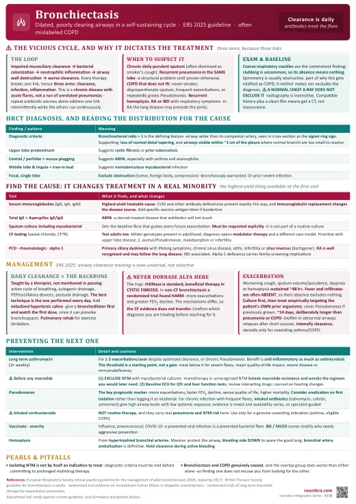
Bronchiectasis is permanent, abnormal dilation of the bronchi, and the reason it persists is a self-reinforcing loop: impaired mucociliary clearance leads to bacterial colonization, colonization drives neutrophilic inflammation, and that inflammation destroys the airway wall, which further impairs clearance. Every effective treatment works by breaking one link in that cycle, which is why the management has three separate arms (clearance, infection, inflammation) rather than one.
The clinical consequence is that this is a chronic disease with acute flares, not a series of unrelated pneumonias. A patient treated only with repeated antibiotic courses, without daily airway clearance, is having one link of the cycle addressed intermittently while the others run continuously.
- Chronic productive cough with daily sputum, often for years and frequently dismissed as smoker's cough or chronic bronchitis. Sputum volume and purulence between exacerbations is the classic tell.
- Recurrent pneumonia in the same anatomic location -a repeated infection in one lobe is a structural problem until proven otherwise.
- COPD that does not fit -a patient labeled COPD who is a never-smoker, has disproportionate sputum production, frequent exacerbations, or repeatedly grows Pseudomonas. Bronchiectasis and COPD also genuinely coexist, and the overlap group does worse than either alone.
- Hemoptysis -bronchial arteries hypertrophy in chronically inflamed airways, so bronchiectasis is a leading cause of recurrent hemoptysis and can produce massive bleeding.
- Rheumatoid arthritis or inflammatory bowel disease with respiratory symptoms -both have well-described associations, and in RA the bronchiectasis may precede the joint disease.
- Crackles are the most common finding, classically coarse and inspiratory; wheeze and rhonchi are also frequent. Clubbing occurs but is uncommon, so its absence means nothing.
- Spirometry typically shows an obstructive pattern, which is part of why the disease gets misfiled under COPD or asthma. Some patients are normal or restrictive, so spirometry neither makes nor excludes the diagnosis.
- Chest radiography is insensitive and a normal film does not exclude bronchiectasis. Tram-track or ring shadows may be visible in advanced disease, but a normal chest X-ray in a patient with a compatible history should prompt a CT, not reassurance.
- High-resolution CT of the chest is the diagnostic standard. The defining feature is bronchial dilation relative to the accompanying pulmonary artery, giving a bronchoarterial ratio greater than 1, seen in cross-section as the signet ring sign.
- Supporting features: lack of normal distal tapering of the airway, and airways visible within about 1 cm of the pleural surface, where normal bronchi are too small to see.
- Read the distribution, because it points at the cause. Upper lobe predominance suggests cystic fibrosis or prior tuberculosis; central or perihilar disease with mucus plugging suggests allergic bronchopulmonary aspergillosis; middle lobe and lingula involvement with tree-in-bud nodules suggests nontuberculous mycobacterial infection; a focal single-lobe pattern raises obstruction (tumor, foreign body) or prior severe infection.
| Test | What it finds and why it matters |
|---|---|
| Serum immunoglobulins (IgG, IgA, IgM) | The single highest-yield treatable cause. Common variable immunodeficiency and other antibody deficiencies present exactly this way, and immunoglobulin replacement changes the disease course. Consider specific antibody titers to pneumococcal and other vaccine antigens if levels are borderline but suspicion is high. |
| Total IgE, Aspergillus-specific IgE and IgG | Screens for allergic bronchopulmonary aspergillosis, which is treated with corticosteroids with or without antifungal therapy rather than with antibiotics. Suspect it particularly with central bronchiectasis, mucus plugging, asthma and eosinophilia. |
| Sputum culture, including mycobacterial and fungal | Establishes the baseline flora that will guide exacerbation treatment. Explicitly request mycobacterial culture -it is not included in routine culture, and NTM must be excluded before starting long-term macrolide therapy. |
| Cystic fibrosis testing (sweat chloride, CFTR genetics) | Test adults, not just children. Milder CFTR genotypes present in adulthood, and a diagnosis opens access to modulator therapy plus a distinct multidisciplinary care model. Prioritize with upper lobe disease, Staphylococcus aureus or Pseudomonas colonization, malabsorption, infertility or a suggestive family history. |
| Primary ciliary dyskinesia evaluation | Consider with lifelong symptoms, chronic sinus disease, otitis media, infertility, or situs inversus (Kartagener syndrome). Nasal nitric oxide is a useful screen where available. |
| Rheumatologic and IBD assessment | Rheumatoid arthritis is a well-recognized association and the lung disease may precede the arthritis. Inflammatory bowel disease is also associated. Check when there are extrapulmonary symptoms. |
| Alpha-1 antitrypsin level | Classically causes emphysema, but bronchiectasis is described, and deficiency has specific implications including family screening. |
| Aspiration and obstruction assessment | For focal disease, exclude an obstructing lesion (tumor, foreign body, extrinsic compression) -bronchoscopy is warranted. For dependent-segment disease, consider recurrent aspiration and swallowing evaluation. |
- Pseudomonas aeruginosa colonization is the marker that most consistently tracks with worse outcomes -more exacerbations, worse quality of life, greater lung function decline and higher mortality. Growing it for the first time should prompt consideration of eradication rather than being logged as an incidental result.
- Validated severity scores (the Bronchiectasis Severity Index and FACED) combine age, body mass index, lung function, radiographic extent, exacerbation history and Pseudomonas status to stratify risk. Their practical use is to identify patients who need more aggressive preventive therapy and closer follow-up.
- Exacerbation frequency is itself prognostic, which is why three or more exacerbations a year is the threshold that triggers escalation to long-term suppressive therapy.
- Daily airway clearance for anyone with chronic productive cough, taught by a respiratory therapist or physiotherapist rather than described in passing. Techniques include the active cycle of breathing, autogenic drainage, positive expiratory pressure devices, oscillatory devices and postural drainage. The best technique is the one the patient will actually perform every day.
- Nebulized hypertonic saline is used as a mucoactive agent to improve sputum clearance. Give a bronchodilator first and monitor the first dose, since hypertonic saline can provoke bronchospasm.
- Pulmonary rehabilitation for patients with exercise limitation -it improves exercise capacity and quality of life, and is underused in this population.
- Definition: worsening of several cardinal symptoms -cough, sputum volume or purulence, breathlessness, fatigue, hemoptysis- sustained for about 48 hours or more and prompting a change in treatment. Fever and infiltrates are often absent, so their absence does not rule out an exacerbation.
- Send a sputum culture before starting antibiotics, then begin empiric therapy without waiting. The culture guides the next course even if this one is empiric.
- Target the patient's own prior organisms -this is the value of having baseline cultures. Cover Pseudomonas empirically in anyone with prior Pseudomonas, using an agent such as ciprofloxacin orally or an antipseudomonal beta-lactam intravenously for more severe illness.
- Treat for about 14 days, which is deliberately longer than for pneumonia or a COPD exacerbation. The rationale is the structural abnormality and biofilm-associated organisms in dilated airways: shorter courses in this population are associated with earlier relapse.
- Intensify airway clearance during the exacerbation rather than pausing it. Reserve systemic corticosteroids for coexisting asthma or COPD -they are not routine treatment for a bronchiectasis exacerbation.
- Long-term macrolide therapy (typically azithromycin three times weekly) for patients with three or more exacerbations a year despite optimized clearance, or for those chronically colonized with Pseudomonas. The benefit is anti-inflammatory and immunomodulatory as much as antimicrobial, and randomized trials (EMBRACE, 2012, BAT, 2013, BLESS, 2013) show meaningful reductions in exacerbation frequency.
- The three-per-year threshold is a starting point, not a hard gate. Treat below it when exacerbations have been severe, when quality of life is substantially affected, when the bronchiectasis is more severe, or when there is a relevant comorbidity such as immunodeficiency -the count is a proxy for risk, and these features raise the risk at any given count.
- ⚠ Two things must be checked before starting a macrolide. First, exclude nontuberculous mycobacterial infection with mycobacterial cultures, because macrolide monotherapy in unrecognized NTM breeds macrolide resistance and compromises the regimen that would later be needed to treat it. Second, check a baseline ECG for QT prolongation and baseline liver function tests, and review interacting drugs. Counsel about hearing changes.
- Inhaled antibiotics (such as nebulized tobramycin, colistin or aztreonam) are used for chronic Pseudomonas infection with frequent exacerbations, delivering high airway concentrations with limited systemic exposure. Evidence is mixed across trials and availability varies, so this is a specialist-guided decision.
- Pseudomonas eradication may be attempted on first isolation, given how consistently chronic colonization tracks with worse outcomes.
- Vaccinate -annual influenza and pneumococcal vaccination, plus COVID-19 and other age-appropriate immunizations. A prevented viral infection is a prevented bacterial exacerbation in this population.
- Inhaled corticosteroids are not routine therapy for bronchiectasis, and carry a real pneumonia and NTM risk in this group. Use them for a genuine coexisting indication such as asthma or eligible COPD, not for the bronchiectasis itself.
- Hemoptysis arises from hypertrophied bronchial arteries. Massive hemoptysis is managed with airway protection, positioning the bleeding side down to protect the good lung, and bronchial artery embolization as the definitive intervention. Withhold airway clearance during active bleeding.
- Nontuberculous mycobacterial infection can be both cause and consequence, and treatment requires prolonged multidrug therapy started only when criteria are met -isolating an organism is not by itself an indication to treat.
- Refer to pulmonology for frequent exacerbations, Pseudomonas or NTM isolation, rapid lung function decline, massive hemoptysis, or when the etiologic workup identifies a cause needing specific therapy. Surgical resection is reserved for focal disease with refractory symptoms or bleeding.
"Mr. R is a 62-year-old never-smoker carried as COPD for six years, presenting with five days of increased sputum volume and purulence plus worsening dyspnea, without fever. He reports daily productive cough at baseline and three courses of antibiotics in the past year. Exam shows coarse inspiratory crackles at both bases and no clubbing. Prior sputum grew Pseudomonas. HRCT shows lower lobe bronchial dilation with a bronchoarterial ratio above 1 and loss of tapering, consistent with bronchiectasis. I think this is a bronchiectasis exacerbation, and the COPD label was probably wrong from the start given he has never smoked and produces sputum daily. I have sent sputum culture including mycobacterial culture and started ciprofloxacin to cover his prior Pseudomonas, planning 14 days. I have asked respiratory therapy to start airway clearance today and teach a technique for home. For the underlying cause I have sent immunoglobulins, total IgE with Aspergillus-specific IgE and IgG, and CFTR testing given the never-smoker history and Pseudomonas. If his mycobacterial cultures are negative and he continues to exacerbate, he would be a candidate for long-term azithromycin, and I would check a QTc before starting."
- Is airway clearance actually happening? Confirm it is ordered, taught and being performed, not just documented once.
- Does the antibiotic match prior cultures, and has a culture been sent this admission?
- Has the etiologic workup been sent if this is a new diagnosis, or has it been assumed idiopathic without testing?
- Sputum volume, purulence and oxygen requirement trended day to day as the response measure.
- Any hemoptysis? Quantify it, and hold airway clearance if there is active bleeding.
- Discharge plan: full antibiotic duration, a written clearance regimen, vaccination status, pulmonary follow-up, and an explicit plan for what to do at the next flare.
| Decision | Approach and why |
|---|---|
| Suspect it | Chronic daily purulent sputum, recurrent same-lobe pneumonia, a never-smoker labeled COPD, or recurrent hemoptysis. A normal chest X-ray does not exclude it -radiography is insensitive. |
| Confirm it | HRCT: bronchoarterial ratio > 1 (signet ring), lack of distal tapering, airways within 1 cm of the pleura. Distribution hints at cause: upper lobe (CF, prior TB), central with plugging (ABPA), middle lobe and lingula (NTM), focal (obstruction). |
| Find the cause | Immunoglobulins, total IgE with Aspergillus serology, sputum culture including mycobacteria, CF testing in adults too, PCD evaluation, rheumatologic assessment, alpha-1 antitrypsin, bronchoscopy for focal disease. A cause is found in about half and changes management in a meaningful minority. |
| Daily backbone | Airway clearance every day, taught properly, plus nebulized hypertonic saline (bronchodilator first, watch the first dose) and pulmonary rehabilitation. Never dornase alfa -harmful here, beneficial in CF. |
| Treat a flare | Culture first, then empiric therapy targeting the patient's own prior organisms; cover Pseudomonas if previously grown. About 14 days, longer than pneumonia, because of biofilm in structurally abnormal airways. Intensify clearance; steroids only for coexisting asthma or COPD. |
| Prevent the next one | Long-term azithromycin if ≥ 3 exacerbations a year -but exclude NTM first and check a baseline QTc. Inhaled antibiotics for chronic Pseudomonas. Vaccinate. Inhaled corticosteroids are not routine and carry pneumonia and NTM risk. |
| Watch for | Pseudomonas colonization as the key prognostic marker, hemoptysis from hypertrophied bronchial arteries (embolization is definitive), and NTM as both cause and consequence. |
- Chase the cause. Stopping at "idiopathic" without testing denies a treatable minority the immunoglobulin replacement, ABPA therapy or CF care that would change their course.
- Clearance is the treatment; antibiotics treat the flare. The vicious cycle runs daily, so intervention has to be daily.
- Do not import cystic fibrosis therapy wholesale. Dornase alfa is the specific trap -helpful in CF, harmful here.