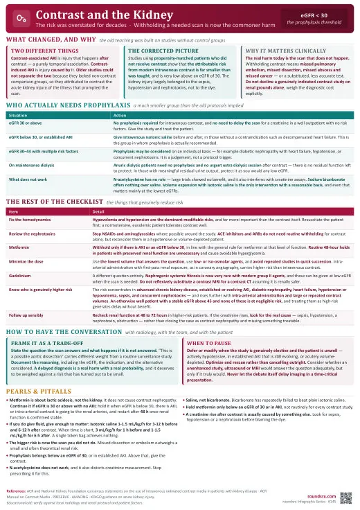
Contrast-Induced Nephropathy
| Risk Factor | Details |
|---|---|
| CKD (eGFR < 30) | #1 risk factor. Risk is very low with eGFR > 45. |
| Diabetes + CKD | Combined = highest risk. Diabetes alone (without CKD) is NOT a significant risk factor. |
| Volume depletion | Dehydration concentrates contrast in kidneys → direct tubular toxicity |
| High contrast volume | Risk proportional to volume. Minimize contrast used. |
| Nephrotoxic medications | NSAIDs, aminoglycosides, ACEi/ARBs (hold if possible day of contrast) |
| Heart failure | Reduced renal perfusion |
- Direct tubular toxicity, osmotic injury to renal tubular epithelial cells from hyperosmolar contrast
- Renal vasoconstriction → medullary ischemia, outer medulla is a vulnerable watershed zone with baseline low oxygen tension; contrast exacerbates ischemia
- Reactive oxygen species (ROS), contrast generates free radicals → oxidative injury to tubular cells
- Increased viscosity, contrast increases tubular fluid viscosity → sluggish flow → prolonged tubular exposure to toxin
- Result: Acute tubular necrosis (ATN) pattern, muddy brown granular casts, elevated FeNa
| Route | Risk | Notes |
|---|---|---|
| IV contrast (CT scans) | Very low risk of true CIN, even in CKD | Multiple large propensity-matched studies show minimal additional AKI risk beyond what would occur without contrast. Do NOT withhold indicated CT scans. |
| Intra-arterial contrast (cardiac cath, angiography) | HIGHER risk | Contrast delivered directly to renal arteries at high concentration. This is where most true CIN occurs. Volume of contrast is the key modifiable risk factor. |
- IV isotonic saline -1 mL/kg/hr for 6–12h before AND 6–12h after contrast. #1 proven prevention.
- Minimize contrast volume -use lowest effective dose
- Hold nephrotoxins -NSAIDs, aminoglycosides. Consider holding ACEi/ARB day of contrast.
- Metformin: Updated (ACR 2022): If eGFR ≥ 45, no need to hold. If eGFR 30–44, hold day of contrast, resume 48h later if Cr stable. eGFR < 30, hold regardless. Risk is lactic acidosis if contrast causes AKI → metformin accumulates. Old blanket "hold metformin for all contrast" is outdated.
- Use low-osmolar or iso-osmolar contrast
- N-acetylcysteine -no longer recommended (multiple trials show no benefit)
- Avoid repeat contrast within 48–72h if possible
| eGFR | Risk Level | Pre-Procedure Protocol |
|---|---|---|
| eGFR > 45 | Low risk | Routine hydration. No special precautions needed. |
| eGFR 30–45 | Moderate risk | IV NS 1 mL/kg/hr × 6–12h before contrast |
| eGFR < 30 | High risk | IV NS 1 mL/kg/hr × 6–12h before AND after contrast. Minimize contrast volume. Consider alternative imaging if appropriate. |
- Do NOT delay emergent imaging for hydration. In life-threatening situations (PE, aortic dissection, stroke, trauma), proceed with contrast CT immediately.
- If time allows: Give a bolus of NS 3 mL/kg over 1h before contrast, then continue hydration post-procedure.
- CIN, if it occurs, is usually self-limited. A missed diagnosis is not.
- NSAIDs, hold before and after contrast
- Aminoglycosides, hold if possible
- ACEi/ARB, consider holding day of procedure (controversial but reasonable in high-risk patients)
- Baseline creatinine + eGFR -assess risk
- BMP at 48–72h post-contrast -check for Cr rise
- Urinalysis -muddy brown granular casts (ATN pattern)
- Urine sodium -may be elevated (tubular injury)
- Assess volume status -dehydration increases risk
| Intervention | Details | Evidence |
|---|---|---|
| IV NS | 1 mL/kg/hr × 6–12h pre- and post-contrast | Best evidence for prevention. Only proven intervention. |
| IV NaHCO₃ | 3 mL/kg/hr × 1h pre, then 1 mL/kg/hr × 6h post | PRESERVE, 2018 showed NOT superior to NS. Use NS instead. |
| N-Acetylcysteine (Mucomyst) | 600–1200 mg PO BID × 2 days | No longer recommended. ACT, 2011 and PRESERVE, 2018 showed no benefit. |
| Hold metformin | Hold 48h AFTER contrast | Prevents lactic acidosis if AKI develops. Resume when Cr stable. |
Patient: 72M with CKD stage 3b (eGFR 38), diabetes. Needs CT abdomen/pelvis for suspected diverticular abscess. Team wants to avoid contrast.
Recommendation: Proceed with contrast CT.
- eGFR 38 = low-moderate risk for CIN
- IV NS 1 mL/kg/hr × 6h before and after contrast
- Hold metformin 48h after contrast
- Missing an abscess that needs drainage is worse than CIN risk
Key lesson: Do not withhold indicated contrast imaging in CKD patients. Hydrate appropriately and proceed.
Patient: 55F post-cardiac catheterization (200 mL contrast), eGFR 25, diabetes. Cr was 2.1 pre-cath, now 2.8 at 48h. UOP decreasing.
Diagnosis: Contrast-associated AKI (intra-arterial route, high risk).
Management:
- Aggressive IV hydration (monitor for volume overload)
- Hold all nephrotoxins
- Monitor Cr daily
- Most cases self-resolve in 7–14 days
- Dialysis rarely needed
Key lesson: Intra-arterial contrast (cardiac cath) carries higher CIN risk than IV contrast. High contrast volume + low baseline eGFR = highest risk combination.
Patient: 40F in ED with acute dyspnea, tachycardia, pleuritic chest pain. Wells score 6 (PE likely). eGFR unknown but Cr was 1.8 last year.
Intern asks: Should we wait for today's Cr before ordering CT-PE?
Answer: NO. Emergent CT-PE now.
- A missed PE can be fatal
- CIN risk is low with IV contrast
- Give concurrent NS bolus while patient is in CT
- Do NOT delay life-saving imaging for CIN prevention
Key lesson: In emergent situations, the risk of a missed diagnosis always outweighs the risk of CIN. Proceed with imaging and hydrate concurrently.