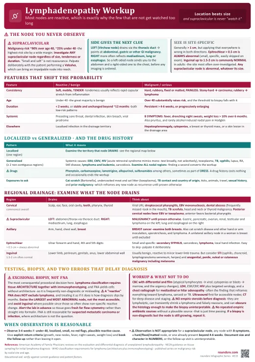
Lymphadenopathy Workup
- Greater than 1 cm is the usual working threshold, but applying it everywhere is wrong in both directions.
- An epitrochlear node above about 0.5 cm is always abnormal and warrants explanation -it is a small, specific finding that is easy to skip on examination.
- Inguinal nodes up to about 1.5 to 2 cm are commonly normal in adults, particularly in people who go barefoot or have recurrent lower limb minor trauma, so this is the site most likely to be over-investigated.
- Any supraclavicular node is abnormal, whatever its size.
- Localized means a single region, and prompts you to examine the territory that node drains for the cause -scalp and dentition for cervical nodes, the arm and hand for axillary, the genitals, perineum and lower limb for inguinal.
- Generalized means two or more non-contiguous regions, and shifts the thinking toward systemic causes: infection (EBV, CMV, HIV, toxoplasmosis, tuberculosis, syphilis), autoimmune disease (lupus, rheumatoid arthritis, Still disease), malignancy (lymphoma, leukemia), sarcoidosis, and drugs.
- ⚠ Do not forget medications. Phenytoin, carbamazepine, lamotrigine, allopurinol and sulfonamides among others can cause lymphadenopathy, sometimes as part of a drug reaction with eosinophilia and systemic symptoms. A drug history costs nothing and occasionally ends the workup.
| Feature | Suggests reactive or benign | Suggests malignant or serious |
|---|---|---|
| Consistency | Soft, mobile, tender -tenderness usually reflects rapid capsular stretch from inflammation | Hard, rubbery, fixed or matted, painless. Stony-hard suggests carcinoma; rubbery suggests lymphoma |
| Age | Under 40, where the great majority of lymphadenopathy is benign | Over 40 substantially raises malignancy risk, and the threshold to biopsy falls accordingly |
| Duration | Under 2 weeks, or stable and unchanged beyond about 12 months -both are low-risk patterns | Persistent beyond 4 to 6 weeks, or progressively enlarging |
| Systemic features | Preceding sore throat, dental infection, local skin breach, or a clear viral prodrome | B symptoms: fever, drenching night sweats, unintentional weight loss (classically more than 10% over 6 months). Also pruritus and, rarely, alcohol-induced nodal pain in Hodgkin lymphoma |
| Extranodal findings | Localized infection in the drainage territory | Hepatosplenomegaly, cytopenias, a breast or thyroid mass, or a skin lesion in the drainage area |
- Exposures: cat scratch or bite (Bartonella), undercooked meat and cat litter (toxoplasmosis), tuberculosis contact and country of origin, tick bites, farm and animal contact, travel.
- Sexual history -acute HIV presents as a mononucleosis-like illness with generalized lymphadenopathy, and syphilis is a classic mimic; both are missed by not asking.
- Occupational and hobby exposures, including silica and beryllium.
- Full medication review as above, and any recent vaccination.
- Prior malignancy, which reframes any new node as possible recurrence until proven otherwise.
- Examine ALL nodal regions, not just the one the patient noticed -cervical, supraclavicular, axillary, epitrochlear, inguinal- because finding a second non-contiguous region converts this to generalized lymphadenopathy and changes the workup entirely.
- Record size, consistency, mobility, tenderness and whether nodes are matted, since these are the features that will be compared at follow-up. "Enlarged node" documented without measurement makes the next visit uninterpretable.
- Examine the drainage territory and the liver and spleen. Inspect skin, scalp and oral cavity in cervical presentations, and breasts in axillary ones.
- Feel for a supraclavicular node deliberately, with the patient performing a Valsalva, which can bring an otherwise impalpable node up into reach.
- Complete blood count with differential and film -atypical lymphocytes point at a viral cause; cytopenias or blasts point at marrow involvement and change the urgency completely.
- LDH, elevated with high cell turnover in lymphoma, and ESR or CRP as non-specific supports.
- Targeted serology and testing based on the history: HIV (test broadly, not selectively), EBV and CMV, toxoplasma, Bartonella, syphilis, and tuberculosis testing where the exposure history or presentation fits.
- Chest radiograph to look for mediastinal or hilar adenopathy, which can be the finding that reframes the whole picture toward lymphoma, sarcoidosis or tuberculosis.
- Ultrasound is the useful first imaging test for accessible nodes, describing size, shape and internal architecture. CT for deep or mediastinal disease, and to map extent once malignancy is suspected.
- ⚠ Do not give empiric corticosteroids before a diagnosis is established. Steroids are lympholytic and can shrink a lymphoma temporarily, which is falsely reassuring, and they can obscure the histology enough to make the eventual biopsy uninterpretable. If a node is worrying enough to want to treat it, it is worrying enough to biopsy first.
- Do not run repeated courses of empiric antibiotics for a persistent node. A short course is reasonable when there is a plausible bacterial source; a second and third course in the absence of one is simply time passing while a diagnosis waits.
- If the biopsy is non-diagnostic but suspicion remains, repeat it. A "reactive" result from a node that is still enlarging has not answered the question.
"Mr. P is a 58-year-old man with a 6-week history of a painless left supraclavicular node, now about 2 cm, hard and fixed. He has lost around 7 kg over 4 months and describes drenching night sweats, so he has B symptoms. This is the highest-risk presentation in lymphadenopathy: a supraclavicular node at his age carries roughly a 90% malignancy risk, and being on the left it raises abdominal or gastrointestinal primaries via thoracic duct drainage, so I have examined the abdomen carefully and will image it. On full nodal examination I found no second non-contiguous region, and there is no hepatosplenomegaly. I have sent a CBC with differential and film, LDH, ESR, HIV and a chest radiograph, and requested CT of chest, abdomen and pelvis. The key management point is that this needs an excisional biopsy, not an FNA: if this is lymphoma, classification requires architecture as well as immunophenotyping, and a reactive FNA result would not exclude it. I have discussed with surgery and alerted pathology so the specimen goes fresh for flow cytometry. I have specifically not started steroids or empiric antibiotics, because steroids could shrink a lymphoma transiently and render the histology uninterpretable."
- Is it supraclavicular? If yes, investigate now regardless of size or symptoms.
- All nodal regions examined, plus liver and spleen, and the drainage territory of the affected node.
- Size and character documented in numbers, so follow-up is interpretable.
- B symptoms asked about explicitly -fever, night sweats, weight loss.
- Drug list and exposure history reviewed, including sexual history and tuberculosis risk.
- If observing: 3 to 4 weeks, explicit return criteria given, follow-up booked rather than left open.
- If biopsying: excisional, largest and most abnormal node, avoid inguinal, and warn the laboratory so tissue arrives fresh.
| Point | Detail |
|---|---|
| ⚠ Location first | Supraclavicular = ~90% malignancy over 40, ~25% under 40. Investigate at ANY size. LEFT (Virchow) = abdominal/GI via thoracic duct; RIGHT = mediastinum, lung, esophagus. Never observe one. |
| Size is site-specific | Generally > 1 cm. But epitrochlear > 0.5 cm is always abnormal, inguinal up to 1.5-2 cm is often normal, and any supraclavicular node is abnormal. |
| Localized vs generalized | Generalized = 2+ non-contiguous regions → systemic causes (EBV, CMV, HIV, toxoplasma, TB, syphilis, lupus, lymphoma, sarcoid, drugs). Localized → examine the drainage territory. |
| Character | Benign: soft, mobile, tender, < 2 weeks or stable > 12 months. Worrying: hard, fixed, matted, painless, > 4-6 weeks or growing, age > 40. Stony-hard suggests carcinoma; rubbery suggests lymphoma. |
| B symptoms | Fever, drenching night sweats, weight loss > 10% over 6 months. Their presence removes observation from the options. |
| Drugs | Phenytoin, carbamazepine, lamotrigine, allopurinol, sulfonamides -sometimes as DRESS. A drug history occasionally ends the workup. |
| Initial tests | CBC with differential and film, LDH, ESR/CRP, HIV plus targeted serology, and a chest radiograph for mediastinal or hilar disease. Ultrasound for accessible nodes, CT for deep disease and staging. |
| ⚠ Biopsy | EXCISIONAL, not FNA, when lymphoma is possible -classification needs architecture + immunophenotyping, and a "reactive" FNA does not exclude lymphoma. Take the largest and most abnormal node, avoid inguinal, and alert the lab so tissue goes fresh for flow cytometry. |
| ⚠ Do not | No empiric steroids before diagnosis -lympholytic, transiently shrink lymphoma, and can render histology uninterpretable. No repeated empiric antibiotic courses without a plausible source. Repeat a non-diagnostic biopsy if suspicion persists. |
| Observation | Reasonable for 3-4 weeks if under 40, localized, small, no red flags, plausible reactive cause -with explicit return criteria and a booked follow-up. |
- Supraclavicular is never "let's watch it." Location beats size, tenderness and duration as a risk signal.
- Excise, do not aspirate, when lymphoma is on the list. A reactive FNA answers nothing.
- No steroids before tissue. Treating the node you have not diagnosed can destroy the diagnosis.