Acute Mesenteric Ischemia
- Serial abdominal exams q2-4h
- Lactate q4-6h
- UOP
- Repeat CTA if change
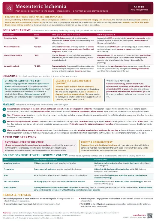
| Type | % of AMI | Mechanism | Key Clue |
|---|---|---|---|
| Arterial embolism (SMA) (~50%) | Most common | Embolus from heart (Afib, LV thrombus, valvular) lodges in SMA | Sudden onset severe pain. Afib is the #1 risk factor. Pain out of proportion to exam. |
| Arterial thrombosis (~25%) | Thrombosis at atherosclerotic plaque (usually SMA origin) | History of chronic mesenteric ischemia (postprandial pain, food fear, weight loss) → acute event. | |
| Mesenteric venous thrombosis (~10%) | SMV thrombosis → venous congestion → mucosal ischemia | More insidious onset (days). Risk: hypercoagulable states, portal HTN, recent surgery, OCP use. | |
| Non-occlusive (NOMI) (~15%) | Splanchnic vasoconstriction in low-flow states | ICU patients on vasopressors, post-cardiac surgery, hemodialysis, shock. No clot -vasospasm. |
- "Pain out of proportion to exam" -severe abdominal pain with a soft, non-tender abdomen (early). The exam catches up later (peritonitis = bowel already dead).
- Nausea, vomiting, bloody diarrhea (late -mucosal sloughing)
- Elevated lactate (often markedly, > 4–5) -but normal lactate does NOT rule it out early
- Leukocytosis, metabolic acidosis, elevated LDH
- Risk factors: Afib, recent MI, CHF, PVD, vasopressor use, hypercoagulable state
| Test | Findings |
|---|---|
| CT angiography (CTA) TEST OF CHOICE | Sensitivity ~94%, specificity ~95% with multidetector CT. Shows arterial/venous filling defects, bowel wall thickening, pneumatosis (gas in bowel wall = necrosis), portal venous gas, free fluid. |
| Lactate | Elevated (often > 4). But normal lactate does not exclude early AMI. Trend is more useful than single value. |
| Plain X-ray | Late findings: pneumatosis intestinalis, portal venous gas, free air (perforation). Normal X-ray does not rule out AMI. |
| Test | What You Are Looking For | Why, and Where It Fails |
|---|---|---|
| CT angiography TEST OF CHOICE | Arterial and portal venous phases. Sensitivity ~94%, specificity ~95%. Look for an SMA filling defect, bowel wall thickening, absent wall enhancement, pneumatosis and portal venous gas. | ⚠ Do NOT give positive oral contrast. It fills the lumen and obscures the very bowel wall you need to assess for enhancement, and waiting for the patient to drink it wastes the time that decides the outcome. ⚠ Do not delay for the creatinine either: the risk of a missed infarction dwarfs the risk of contrast nephropathy. |
| Lactate CANNOT RULE OUT | Often above 4, and rising on serial draws. | ⚠ A normal lactate does not exclude acute mesenteric ischemia, and this is the error that kills. Lactate reflects advanced injury and systemic hypoperfusion, not early ischemia, so by the time it climbs the bowel is often already infarcted. It is a marker of how late you are, never a screening test. The trend is worth more than any single value. |
| CBC, BMP, ABG | Leukocytosis, metabolic acidosis with an anion gap, AKI from hypovolemia. | Consistent but entirely non-specific, and all of them can be normal early. Their value is in tracking deterioration, not in making or excluding the diagnosis. Treat a normal panel in a convincing story as reassurance about nothing. |
| Plain radiograph | Pneumatosis intestinalis, portal venous gas, free air. | These are late findings that mean bowel has already died. A normal film rules out nothing. Its only real use is looking for free air or an alternative diagnosis such as obstruction. |
| ECG and echocardiogram | Atrial fibrillation, recent myocardial infarction, left ventricular thrombus, valvular disease. | Finds the embolic source behind roughly half of all cases. It does not change the emergency, but it determines anticoagulation afterwards and whether the other vascular beds are at risk. |
| Drug | Dose | Route | Why This, and What to Watch |
|---|---|---|---|
| Heparin ALL SUBTYPES | 80 U/kg bolus, then 18 U/kg/h | IV | Stops clot propagation while the plan is made, and it is the definitive treatment on its own for mesenteric venous thrombosis, which usually does not need an operation. ⚠ Discuss the bolus with surgery first if laparotomy is imminent. |
| Fluid resuscitation | Titrated to perfusion | IV | These patients are profoundly volume-depleted from third-spacing into ischemic bowel, and the deficit is usually larger than it looks. Restoring volume is also the treatment for NOMI, where the whole problem is splanchnic hypoperfusion. |
| Piperacillin-tazobactam | 4.5 g q6h | IV | Ischemic bowel loses its mucosal barrier and translocates gut flora into the bloodstream, so cover enteric Gram-negatives and anaerobes early rather than waiting for peritonitis to be obvious. |
| Papaverine NOMI ONLY | 30–60 mg/h | Intra-arterial | A direct vasodilator infused into the SMA to break the splanchnic vasospasm that IS the disease in NOMI. Pointless in embolic or thrombotic disease, where the problem is a clot and not tone. |
| Reconsider the vasopressors IN NOMI | – | – | ⚠ In NOMI the vasoconstrictors keeping the blood pressure up are also driving the bowel ischemia. Where the hemodynamics allow, optimize volume and cardiac output so the alpha-agonist dose can come down, and review digoxin, which causes splanchnic vasoconstriction in its own right. This is a genuine trade-off to make with the intensivist, not a unilateral change. |
Patient: 72F with Afib (not on anticoagulation). Sudden severe periumbilical pain × 4h, nausea, bloody diarrhea. HR 110, BP 95/60. Abdomen: diffuse tenderness but soft, no rebound. Lactate 5.2, WBC 19K.
Key findings: Pain out of proportion to exam, classic for early mesenteric ischemia. Afib without anticoagulation = #1 risk for SMA embolism. Elevated lactate confirms tissue ischemia.
Management:
- Emergent CT angiography (CTA), expect abrupt SMA cutoff at mid-vessel (emboli lodge at branch points)
- IV heparin bolus + drip (prevent clot propagation)
- Emergent surgery consult for SMA embolectomy + bowel viability assessment
- Broad-spectrum antibiotics (piperacillin-tazobactam), bacterial translocation through ischemic bowel wall
- Aggressive IVF resuscitation, third-spacing and lactic acidosis
Teaching point: The golden window for mesenteric ischemia is before peritoneal signs develop. Once the abdomen becomes rigid, bowel necrosis is likely irreversible. A soft abdomen with severe pain = act now.
Patient: 68M in ICU on norepinephrine 0.3 mcg/kg/min for cardiogenic shock post-MI. Develops abdominal distension, bloody NG output, rising lactate 3.2 → 7.8. WBC 24K.
Key findings: Non-occlusive mesenteric ischemia, splanchnic vasoconstriction from shock + vasopressors. No embolic source. CTA may show patent but narrowed mesenteric vessels with poor bowel wall enhancement.
Management:
- Optimize cardiac output (the root cause), inotropes, IABP, reduce vasopressors if possible
- CTA to confirm NOMI (patent vessels but poor bowel enhancement) and exclude occlusive disease
- If confirmed NOMI: papaverine infusion via SMA catheter (direct splanchnic vasodilator) if available
- NPO, NGT decompression, serial abdominal exams q4h
- Surgery consult, if peritoneal signs develop, exploratory laparotomy for bowel viability assessment
Teaching point: NOMI accounts for ~20% of mesenteric ischemia and has the highest mortality (60-80%) because the underlying cause (shock) is often difficult to reverse. The treatment is hemodynamic optimization, not anticoagulation or surgery.
Patient: 65F smoker with PVD. Postprandial epigastric pain × 6 months, occurring 15-30 min after eating, lasting 1-2h. "Food fear", eating less, lost 20 lb. CTA: > 70% stenosis of SMA and celiac artery.
Key findings: Classic chronic mesenteric ischemia triad: postprandial pain + food avoidance + weight loss. Requires ≥ 2 of 3 mesenteric vessels to be stenotic for symptoms (collateral supply compensates for single vessel disease).
Management:
- Mesenteric angiography with possible stenting of SMA (first-line for chronic mesenteric ischemia)
- Surgical bypass (SMA bypass graft) if endovascular approach fails or not feasible
- Nutritional optimization, small frequent meals, supplemental nutrition pre-procedure
- Aggressive atherosclerotic risk factor management (statin, ASA, smoking cessation)
- Urgent revascularization, risk of acute-on-chronic ischemia (bowel infarction) is 30-50% if untreated
Teaching point: Chronic mesenteric ischemia is the "angina of the gut." The diagnosis is often delayed because postprandial pain has a broad differential. Weight loss + food fear + vascular risk factors should trigger CTA of mesenteric vessels.
- Serial abdominal exams q2-4h
- Lactate q4-6h
- UOP
- Repeat CTA if change
Text version
- Reassured by benign abdominal exam (early ischemia = soft abdomen + severe pain)
- Waiting for lactate to elevate (late marker)
- Not thinking about it (must suspect to diagnose)
- Delaying surgical consult