Nephrolithiasis
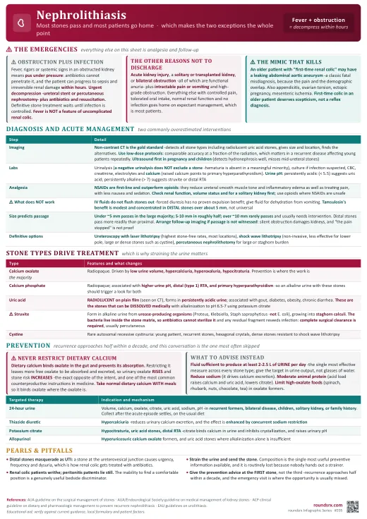
Most stones pass and most patients go home, which makes the two exceptions the whole point: an obstructing stone with infection is a urologic emergency needing decompression within hours, and a stone in a solitary or transplanted kidney is functional anuria. Everything else is analgesia, hydration and expectant management -plus the prevention conversation that almost never happens, in a disease with a recurrence rate approaching half within a decade.
🔍 Overview
Presentation
- Sudden severe colicky flank pain radiating to the groin, classically with the patient unable to find a comfortable position -in contrast to peritonitis, where patients lie still. Nausea and vomiting are common.
- Hematuria (usually microscopic) is present in most but absent in a meaningful minority, so a negative urinalysis does not exclude a stone.
- Pain location tracks stone position: flank pain with proximal ureteral stones, migrating toward the groin as it descends, and urinary urgency, frequency and dysuria when the stone reaches the ureterovesical junction -which is why distal stones get misdiagnosed as UTI.
- Fever is not part of uncomplicated renal colic. Fever plus obstruction is the emergency below.
Stone Types (they drive prevention)
- Calcium oxalate -the majority. Radiopaque. Driven by low urine volume, hypercalciuria, hyperoxaluria, hypocitraturia.
- Calcium phosphate -radiopaque; associated with higher urine pH, distal (type 1) renal tubular acidosis, and primary hyperparathyroidism.
- Uric acid -radiolucent on plain film (visible on CT), forms in persistently acidic urine, associated with gout, diabetes, obesity and chronic diarrhea. These are the stones that can be dissolved medically with urinary alkalinization.
- Struvite (magnesium ammonium phosphate) -form in alkaline urine from urease-producing organisms (Proteus, Klebsiella, Staphylococcus saprophyticus, not E. coli). They grow into staghorn calculi, and because they are infected stones, complete surgical clearance is required -antibiotics alone will not sterilize them.
- Cystine -rare, autosomal recessive cystinuria, presenting young with recurrent stones and hexagonal crystals.
Obstructing stone plus infection is a urologic emergency. Fever, rigors, or systemic signs in a patient with an obstructed kidney means pus under pressure, which does not respond to antibiotics alone and can progress to sepsis and irreversible renal damage within hours. Urgent decompression -ureteral stent or percutaneous nephrostomy- plus antibiotics and resuscitation. Definitive stone treatment waits until the infection is controlled. This is the single most time-critical decision in the topic.
🧪 Diagnosis
Imaging
- Non-contrast CT of the abdomen and pelvis is the gold standard -detects essentially all stone types including radiolucent uric acid stones, gives size and location, and identifies the alternative diagnoses that matter.
- Use low-dose CT protocols where available: comparable accuracy for stone detection at a fraction of the radiation, which matters in a recurrent disease that often affects young patients repeatedly.
- Ultrasound first in pregnancy and in children, and as a reasonable initial test in young patients with a typical presentation and prior confirmed stones -it detects hydronephrosis reliably but is far less sensitive for the stone itself, especially in the mid-ureter.
- Plain radiography (KUB) has a limited role beyond following a known radiopaque stone.
- Consider the mimics that kill: abdominal aortic aneurysm in an older patient with "first-time renal colic" -this is a classic fatal misdiagnosis- plus appendicitis, ovarian torsion, ectopic pregnancy, and mesenteric ischemia.
Laboratory
- Urinalysis (hematuria, pH, crystals, and signs of infection), urine culture if infection is suspected, CBC, creatinine and electrolytes, and calcium -a raised calcium points to primary hyperparathyroidism.
- Urine pH is informative: persistently acidic (below about 5.5) suggests uric acid stones, while persistently alkaline (above about 7) suggests struvite or distal RTA.
- Strain the urine and send any recovered stone for analysis -stone composition is the single most useful piece of preventive information, and it is routinely lost because nobody strains.
🚨 Acute Management
Analgesia
- NSAIDs are first-line and outperform opioids for renal colic -they reduce ureteral smooth muscle tone and inflammatory edema as well as providing analgesia, with less nausea and sedation. Ketorolac is the usual parenteral choice.
- Check renal function and volume status first: avoid NSAIDs in acute kidney injury, significant CKD, or a solitary kidney, and use opioids there instead.
- Opioids are a rescue, not a default; combine with an antiemetic. IV fluids do not "flush the stone out" and forced diuresis has no proven benefit -give fluid to correct dehydration from vomiting, not as a stone-expulsion strategy.
Who Goes Home, Who Stays
- Discharge with expectant management if pain is controlled on oral medication, the patient can tolerate fluids, renal function is normal and there is no infection -most stones are managed this way.
- Admit and involve urology urgently for: infection with obstruction (the emergency), acute kidney injury, a solitary or transplanted kidney or bilateral obstruction (functional anuria), intractable pain or vomiting, and high-grade obstruction.
- Size predicts passage: stones under about 5 mm pass spontaneously in the large majority, 5-10 mm pass in roughly half, and stones over about 10 mm rarely pass and usually need intervention. Distal stones pass more readily than proximal ones.
Medical Expulsive Therapy and Definitive Treatment
- Alpha-blockers (tamsulosin) for distal ureteral stones larger than about 5 mm -the benefit is modest and concentrated in that subgroup, which is worth stating honestly rather than prescribing it universally. It relaxes ureteral smooth muscle to aid passage.
- Follow-up imaging within a few weeks if the stone has not visibly passed -silent obstruction can damage a kidney without symptoms, and "the pain stopped" does not prove the stone passed.
- Definitive options: ureteroscopy with laser lithotripsy (highest stone-free rates, works for most locations and compositions), shock wave lithotripsy (non-invasive but less effective for lower pole, large, or dense stones such as cystine), and percutaneous nephrolithotomy for large or staghorn burden.
- Uric acid stones can be dissolved with urinary alkalinization to a pH of roughly 6.5-7 using potassium citrate -one of the few stones amenable to medical dissolution, which is why identifying composition matters.
💊 Prevention
Why It Matters
Recurrence approaches half of patients within a decade, and the preventive conversation is the part of this disease most often skipped in a busy emergency department. A first stone earns dietary advice for everyone; recurrent, bilateral or high-risk stone formers earn a metabolic evaluation.
Universal Advice (all stone types)
- Fluid intake sufficient to produce at least 2-2.5 L of urine per day -the single most effective intervention across every stone type, and the one patients most often under-do. Give the target in urine output, not in glasses of water.
- Reduce sodium -high sodium intake drives calcium excretion, so salt restriction lowers urinary calcium.
- Moderate animal protein -an acid load that raises calcium and uric acid excretion and lowers citrate.
- ⚠ Do NOT restrict dietary calcium. Normal dietary calcium binds oxalate in the gut and reduces its absorption, so restricting it paradoxically increases urinary oxalate and stone risk. This is one of the most common and most counterproductive pieces of patient advice in medicine.
- Limit high-oxalate foods (spinach, rhubarb, nuts, chocolate, tea) in calcium oxalate formers, and take calcium with meals so it can bind oxalate where it matters.
Metabolic Evaluation and Targeted Therapy
- 24-hour urine collection (volume, calcium, oxalate, citrate, uric acid, sodium, pH) in recurrent stone formers, bilateral disease, children, patients with a solitary kidney, and those with a family history -done after the acute episode has settled and on the patient's usual diet.
- Thiazide diuretics for hypercalciuria -they reduce urinary calcium excretion, which is the mechanism, and the effect is enhanced by concurrent sodium restriction.
- Potassium citrate for hypocitraturia, uric acid stones and distal RTA -citrate binds calcium in urine and inhibits crystallization, and it raises urinary pH.
- Allopurinol for hyperuricosuric calcium oxalate stone formers and for uric acid stones where alkalinization alone is insufficient.
- Check for primary hyperparathyroidism when calcium is elevated, and consider distal RTA with calcium phosphate stones plus a persistently alkaline urine and a non-anion-gap metabolic acidosis.
🏥 Rounds
Pimp Questions
❓ Why should a patient with calcium oxalate stones NOT be told to restrict dietary calcium?
Because dietary calcium binds oxalate in the gut and prevents its absorption. Restricting calcium leaves more free oxalate to be absorbed and excreted, so urinary oxalate rises and stone risk increases -the opposite of the intended effect. Patients should take normal dietary calcium, ideally with meals so it binds oxalate where the oxalate is. What should be restricted instead is sodium (which drives calcium excretion), excess animal protein, and high-oxalate foods in oxalate stone formers. This is one of the most common and most counterproductive pieces of advice patients receive.
❓ A patient with an obstructing ureteral stone spikes a fever to 38.9°C. What is the next step?
Urgent decompression -ureteral stent or percutaneous nephrostomy- plus IV antibiotics and resuscitation. An obstructed, infected collecting system is pus under pressure: antibiotics cannot penetrate it adequately and the patient can progress to sepsis and irreversible renal damage within hours. Definitive stone treatment is deferred until the infection is controlled -attempting lithotripsy or ureteroscopy on an infected obstructed system risks disseminating infection. This is the most time-critical decision in nephrolithiasis, and fever is not a feature of uncomplicated renal colic.
❓ Which stones are radiolucent on plain film, and why does that matter?
Uric acid stones (and cystine stones are only faintly opaque). They are invisible on KUB but readily seen on non-contrast CT, which is one reason CT is the diagnostic standard. It matters therapeutically too: uric acid stones are the ones that can be dissolved medically with urinary alkalinization to a pH of roughly 6.5-7 using potassium citrate. They form in persistently acidic urine and are associated with gout, diabetes, obesity and chronic diarrhea -so identifying the composition changes the treatment from procedural to medical.
❓ Why do struvite staghorn stones require complete surgical removal rather than antibiotics?
Because struvite stones are infected stones, not stones that happen to be infected. They form when urease-producing organisms (Proteus, Klebsiella, Staphylococcus saprophyticus -notably not E. coli) split urea into ammonia, alkalinizing the urine and precipitating magnesium ammonium phosphate. The bacteria live within the stone matrix, so antibiotics cannot sterilize it, and any residual fragment reseeds infection and regrows the stone. Complete clearance, usually by percutaneous nephrolithotomy, is required, and untreated staghorn calculi risk renal loss and urosepsis.
❓ Do IV fluids help a stone pass, and does tamsulosin work?
Two commonly overestimated interventions. IV fluids do not flush stones out -forced diuresis has no proven benefit for expulsion, and fluid should be given to correct dehydration from vomiting rather than as a stone-passing strategy. Tamsulosin has modest benefit that is concentrated in distal ureteral stones larger than about 5 mm; for small stones, which mostly pass anyway, the added benefit is small. Saying this honestly matters because both interventions are often presented to patients as more powerful than they are. What does predict passage is size and location: under 5 mm passes in the large majority, over 10 mm rarely passes.
Sample Presentation
Mr. Kowalczyk is a 41-year-old man with 4 hours of severe right flank pain radiating to the groin, writhing and unable to get comfortable, with vomiting. Afebrile, HR 96, BP 148/88. Urinalysis: microscopic hematuria, no nitrites or leukocyte esterase. Creatinine normal. Low-dose non-contrast CT: 4 mm stone at the right ureterovesical junction with mild hydronephrosis. This is his second stone in three years.
Key Points: (1) He is afebrile with normal renal function and a small distal stone, so this is outpatient expectant management: NSAIDs first-line (renal function is normal, so ketorolac is appropriate), antiemetics, and oral analgesia at home. A 4 mm distal stone passes spontaneously in the large majority. (2) Give him a strainer and tell him why -stone composition is the most useful preventive information available and it is routinely lost. Arrange follow-up imaging in a few weeks if he does not see it pass, because silent obstruction damages kidneys and "the pain stopped" does not prove passage. (3) This is his second stone, so he earns the prevention conversation now: fluid to produce 2-2.5 L of urine daily, reduced sodium, moderated animal protein, and explicitly do not restrict dietary calcium. A 24-hour urine study after this episode settles will direct targeted therapy.
ED & Clinic Checklist
- Fever and infection screened first -obstruction plus infection is decompression within hours, not antibiotics alone.
- Solitary or transplanted kidney identified -obstruction there is functional anuria.
- AAA considered in the older patient with first-time "renal colic."
- NSAID given first-line unless renal function, volume status or a solitary kidney forbids it.
- Size and location documented -they predict passage and drive disposition.
- Strainer given and stone analysis explained; urine pH noted.
- Follow-up imaging arranged if passage is not witnessed.
- Prevention discussed at the first stone -urine output target, sodium, protein, and the do-not-restrict-calcium point; 24-hour urine for recurrent or high-risk formers.
📋 Summary
Summary
Infection Plus Obstruction Is the Emergency
Fever or systemic signs with an obstructed kidney means pus under pressure, which antibiotics cannot penetrate and which progresses to sepsis and irreversible renal damage within hours. Urgent decompression by stent or nephrostomy, plus antibiotics and resuscitation, with definitive stone treatment deferred until infection is controlled. Fever is not a feature of uncomplicated renal colic.
The Other Reasons Not to Discharge
Acute kidney injury, a solitary or transplanted kidney, bilateral obstruction (all functional anuria), intractable pain or vomiting, and high-grade obstruction. Everything else with controlled pain, tolerated oral intake, normal renal function and no infection goes home on expectant management -which is most patients.
CT Is the Standard, but Use Low Dose
Non-contrast CT detects essentially all stone types including radiolucent uric acid stones and gives size and location. Low-dose protocols have comparable accuracy, which matters in a recurrent disease affecting young patients repeatedly. Ultrasound first in pregnancy and children. Remember that a negative urinalysis does not exclude a stone -hematuria is absent in a meaningful minority.
Do Not Miss the Aneurysm
An older patient with "first-time renal colic" may have a leaking abdominal aortic aneurysm -a classic fatal misdiagnosis, because the pain and the demographic overlap. Also consider appendicitis, ovarian torsion, ectopic pregnancy and mesenteric ischemia. First-time colic in an older patient deserves a moment's scepticism rather than a reflex diagnosis.
NSAIDs Beat Opioids for Renal Colic
NSAIDs are first-line and outperform opioids, reducing ureteral smooth muscle tone and inflammatory edema as well as treating pain, with less nausea and sedation. Check renal function, volume status and for a solitary kidney first, using opioids where NSAIDs are unsafe. IV fluids do not flush stones out -give fluid for dehydration, not as an expulsion strategy.
Size and Location Predict Passage
Under about 5 mm passes in the large majority; 5-10 mm in roughly half; over 10 mm rarely passes and usually needs intervention. Distal stones pass more readily than proximal. Tamsulosin's benefit is modest and concentrated in distal stones over about 5 mm, which is worth saying honestly. Arrange follow-up imaging if passage is not witnessed: silent obstruction damages kidneys, and "the pain stopped" is not proof.
Composition Changes the Plan
Strain the urine and send the stone -it is the most useful preventive information and it is routinely lost. Uric acid stones are radiolucent, form in acidic urine, and can be DISSOLVED with alkalinization to pH 6.5-7. Struvite stones are infected stones from urease-producing organisms (Proteus, Klebsiella, Staph saprophyticus), grow into staghorns, and require complete surgical clearance because the bacteria live inside the stone.
Never Restrict Dietary Calcium
Dietary calcium binds oxalate in the gut; restricting it raises urinary oxalate and increases stone risk -the exact opposite of the intent, and one of the most common counterproductive instructions in medicine. Instead: fluid to make 2-2.5 L of urine daily (the single most effective measure across all stone types), reduce sodium, moderate animal protein, take calcium with meals. Recurrent formers get a 24-hour urine, then thiazides for hypercalciuria, potassium citrate for hypocitraturia and uric acid stones, allopurinol for hyperuricosuria.