Pneumothorax
| Type | Cause | Key Features |
|---|---|---|
| Primary spontaneous (PSP) | Rupture of apical bleb/bulla in otherwise healthy lung | Tall, thin, young male, smoker. Usually small. Low recurrence after first episode (~30%), high after second (~50%). |
| Secondary spontaneous (SSP) | Underlying lung disease (COPD, CF, Pneumocystis, ILD, LAM) | More dangerous -limited pulmonary reserve. Even small PTX can cause significant compromise. Lower threshold for intervention. |
| Traumatic | Blunt/penetrating chest trauma, rib fractures | Chest tube. Evaluate for hemothorax (check pleural Hct). |
| Iatrogenic | Central line (subclavian > IJ), thoracentesis, lung biopsy, positive pressure ventilation | Post-procedure CXR. Small iatrogenic PTX in stable patient → may observe. |
| Tension | One-way valve effect → progressive air trapping → mediastinal shift → ↓ venous return → hemodynamic collapse | CLINICAL DIAGNOSIS: hypotension + JVD + absent breath sounds + tracheal deviation away. Needle decompression BEFORE imaging. 14–16G needle, 2nd ICS, midclavicular line (or 5th ICS, anterior axillary line). |
- Apical subpleural bleb rupture -most common. Tall, thin, young male; smoker (~20× risk).
- COPD / emphysema (bullae rupture) -most common cause of SSP. Older smoker. Even a small PTX is dangerous (no pulmonary reserve).
- Severe asthma -during acute exacerbation. Concomitant pneumomediastinum possible.
- Cystic fibrosis -young patient with recurrent infections and bronchiectasis. PTX is a poor prognostic sign.
- Alpha-1 antitrypsin deficiency -young non-smoker (or modest smoker) with basal-predominant emphysema. Co-existing liver disease.
- Pneumocystis jirovecii pneumonia (PCP) -HIV/AIDS or other immunocompromise. Can cause bilateral PTX. High recurrence.
- Tuberculosis (cavitary) -endemic exposure, weight loss, night sweats, cavitary lesion on imaging.
- Lymphangioleiomyomatosis (LAM) -young woman of reproductive age with recurrent PTX. Tuberous sclerosis association, renal angiomyolipomas. Treat with sirolimus.
- Idiopathic pulmonary fibrosis (IPF) -late-stage disease with honeycombing. PTX heralds poor prognosis.
- Marfan syndrome (FBN1) -tall, arachnodactyly, lens dislocation, aortic root dilation. Apical blebs.
- Ehlers-Danlos (vascular type, COL3A1) -hyperextensible skin, joint laxity. Vessel and organ rupture risk.
- Catamenial pneumothorax (thoracic endometriosis) -right-sided, recurrent, within 24-72h of menses in reproductive-age women. Treat with hormonal suppression plus VATS.
- Lung malignancy (primary or metastatic) -cavitary lesion erodes into pleura. Smoker, weight loss.
- Blunt chest trauma -rib fracture lacerating pleura. MVC, fall, sports injury. Often associated with hemothorax.
- Penetrating trauma -stab wound, gunshot. Open ("sucking") chest wound: cover with three-sided dressing.
- Barotrauma -diving (rapid ascent without exhalation), blast injury. Concomitant air embolism risk.
- Central line placement (subclavian > IJ) -~1-3% rate even with ultrasound guidance. Always get post-line CXR.
- Thoracentesis / pleural drainage -~3% with US guidance, > 10% without.
- CT-guided transthoracic needle biopsy -highest iatrogenic rate (~15-20%). Most resolve with observation alone.
- Mechanical ventilation barotrauma -high PEEP, high tidal volumes, ARDS. Watch for progression to tension PTX, especially in patients with bullous disease.
- CPR -rib fractures, occasionally direct pleural injury. Always check post-resuscitation CXR after prolonged or vigorous CPR.
- Tall thin young male, sudden pleuritic pain at rest: primary spontaneous (apical bleb).
- Recurrent right-sided PTX in reproductive-age woman, around menses: catamenial / thoracic endometriosis.
- Young woman with renal angiomyolipomas, recurrent PTX: LAM (check for tuberous sclerosis).
- PTX + skin fibrofolliculomas + renal tumors: Birt-Hogg-Dubé.
- HIV with bilateral PTX: PCP until proven otherwise.
- Post-procedure PTX in a previously stable patient: recently placed line, biopsy, or thoracentesis.
| Scenario | Management |
|---|---|
| Small PSP (< 2 cm at apex), stable | Observation + high-flow O₂ (accelerates reabsorption 4×). Repeat CXR in 4–6h. Discharge if stable and improving. |
| Large PSP (≥ 2 cm) or symptomatic | Needle aspiration (14–16G, 2nd ICS) → recheck CXR. If re-expands → observe. If fails → chest tube. BTS Guidelines, 2023: aspiration first-line for PSP. |
| SSP -any size | Chest tube (14–28 Fr) connected to water seal or low suction (−20 cmH₂O). These patients have no reserve. Do NOT just observe SSP. |
| Tension PTX | Immediate needle decompression → chest tube. Do not wait for CXR. |
| Recurrent PSP (≥ 2 episodes ipsilateral) | VATS with pleurodesis (mechanical or chemical). Recurrence after first: ~30%. After VATS: < 5%. |
Patient: 24M, tall and thin (6'3", 155 lb), sudden right-sided pleuritic chest pain while at rest. SpO₂ 96%. CXR: 25% right pneumothorax. Hemodynamically stable. No underlying lung disease.
Key findings: Primary spontaneous PTX, rupture of apical subpleural blebs in a classic demographic (tall, thin, young male, smoker). No underlying lung disease. Moderate size (> 2 cm at hilum).
Management:
- Needle aspiration with small-bore catheter (14-16G, 2nd ICS MCL), first-line for large primary spontaneous PTX per BTS guidelines
- If aspiration successful (lung re-expands on repeat CXR): observe 4-6h → discharge with 24-48h follow-up CXR
- If aspiration fails: small-bore chest tube (12-14 Fr) with Heimlich valve or underwater seal
- Supplemental O₂ (increases nitrogen gradient → accelerates pneumothorax reabsorption 4×)
- Smoking cessation counseling (recurrence risk 30% if continues smoking vs 15% if stops)
Teaching point: Needle aspiration is first-line for primary spontaneous PTX, it's as effective as chest tube with less pain, shorter hospital stay, and fewer complications. Large-bore chest tubes are overused for simple pneumothoraces.
Patient: 68M with severe COPD (FEV1 28%). Acute worsening dyspnea. SpO₂ 82% on 2L NC (baseline 90%). CXR: left pneumothorax ~20%. HR 112, BP 108/68.
Key findings: Secondary spontaneous PTX in COPD, even a small PTX is dangerous because of minimal pulmonary reserve. BTS guidelines: ALL secondary PTX > 1 cm or symptomatic need intervention (lower threshold than primary).
Management:
- Small-bore chest tube (12-14 Fr), not aspiration (higher failure rate in secondary PTX)
- Connect to underwater seal with -20 cmH₂O suction
- Admit to monitored bed (not discharge like primary PTX)
- If persistent air leak > 5 days: thoracic surgery consult for VATS pleurodesis
- Careful O₂ titration, target SpO₂ 88-92% (COPD CO₂ retainer risk)
Teaching point: Secondary PTX is always more dangerous than primary, the diseased lung cannot compensate. Threshold for intervention is lower (any symptomatic PTX), and these patients require admission, not outpatient management.
Patient: 28M with third right-sided spontaneous PTX in 2 years. Current episode: 35% PTX, managed with chest tube (resolved in 48h). Previous 2 episodes also required chest tubes.
Key findings: Recurrent ipsilateral PTX, after first episode, recurrence risk is ~30%. After second episode, ~50%. After third, > 80%. Definitive prevention is indicated.
Management:
- VATS (video-assisted thoracoscopic surgery) with blebectomy + mechanical pleurodesis, gold standard for recurrent PTX
- VATS: resect apical blebs + abrade parietal pleura → adhesion formation prevents recurrence (< 5% recurrence after VATS)
- Alternative: chemical pleurodesis via chest tube (talc slurry) if not a surgical candidate
- CT chest to assess bleb distribution before surgery
- Absolute indication for pleurodesis: second ipsilateral PTX, first contralateral PTX (bilateral), occupational risk (pilots, divers)
Teaching point: Pleurodesis after second ipsilateral PTX is standard of care. VATS blebectomy + pleurodesis reduces recurrence from > 50% to < 5%. Pilots and scuba divers should have pleurodesis after FIRST episode due to occupational hazard.
| Test | What You Are Looking For | Why, and Where It Fails |
|---|---|---|
| Upright PA chest radiograph FIRST TEST | A visible visceral pleural line with no lung markings beyond it. Measure the rim of air to size it, using the thresholds in the Management tab. | Fast, available, and adequate for most stable presentations. Upright matters: air collects at the apex, so an upright film puts the abnormality where you are already looking. |
| Supine radiograph EASY TO MISS | Deep sulcus sign: an abnormally deep, lucent costophrenic angle. Also a lucent upper abdomen and a sharply outlined hemidiaphragm or cardiac border. | This is where pneumothoraces get missed. Supine, the air layers anteriorly rather than at the apex, so there is often no pleural line to see. Trauma and ICU patients are filmed supine precisely when the diagnosis matters most. |
| Lung ultrasound BEDSIDE | Absent lung sliding and absent B-lines, with a lung point where sliding starts again. On M-mode the normal seashore pattern becomes a barcode (stratosphere) pattern. | Outperforms the supine film: pooled sensitivity 0.91 and specificity 0.99 against supine radiography in trauma across 13 studies (Cochrane, 2020). Sliding or B-lines present rules out pneumothorax in the zone scanned, and a lung point is essentially diagnostic at 100% specificity, though only 66% sensitive Lichtenstein, 2000. Absent sliding alone is not enough: it also occurs in mainstem intubation, pleural adhesions, ARDS and apnea. |
| CT chest DEFINITIVE | The most sensitive test, and the one that finds occult pneumothorax invisible on a supine film. | Reserve it for the questions a plain film cannot answer: a complex or uncertain picture, suspected underlying lung disease, bleb distribution before surgery, or distinguishing a large bulla from a pneumothorax. That last distinction is the one worth slowing down for, because draining a giant bulla mistaken for a pneumothorax creates a persistent air leak. |
- Pulse oximetry and an ABG if hypoxic or hypercapnic, since a pneumothorax in someone with existing lung disease decompensates far faster than the same size in a healthy chest.
- ECG in a left-sided pneumothorax, which can mimic ischemia with reduced voltages, axis shift and precordial T-wave changes. Recognizing it prevents an unnecessary cardiac workup.
- Ask what caused it, because it changes what happens next. Height, low body mass and smoking point to primary spontaneous; COPD, prior tuberculosis or malignancy point to secondary, which is treated more aggressively; recent central line, thoracentesis, biopsy or positive-pressure ventilation points to iatrogenic.
- Ask about recurrence and about air travel or diving, since a recurrent pneumothorax changes the threshold for definitive surgical management, and diving is a lifelong contraindication after an untreated spontaneous pneumothorax.
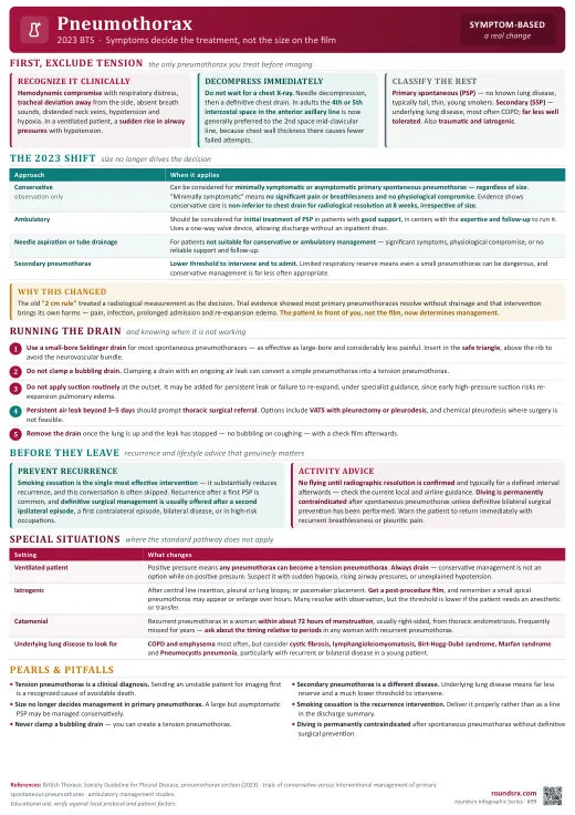
Text version
- Waiting for CXR in tension PTX (clinical diagnosis → decompress)
- Discharging without recurrence counseling (30% after 1st episode)
- Not admitting secondary PTX (these patients decompensate quickly)
- Flying within 2 weeks (risk of re-expansion)