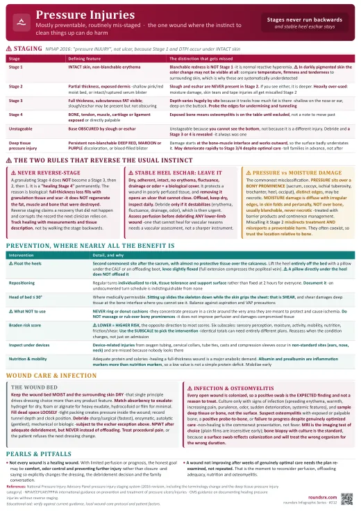
Pressure Injuries
| Stage | Defining feature | The distinction that gets missed |
|---|---|---|
| Stage 1 | INTACT skin with non-blanchable erythema | Blanchable redness is not a Stage 1, it is normal reactive hyperemia. Press it: if it whitens and refills, the microcirculation is intact. In darkly pigmented skin the color change may not be visible at all, so compare temperature, firmness and tenderness against surrounding skin rather than relying on redness, which is why these injuries are systematically underdetected. |
| Stage 2 | Partial-thickness loss with exposed dermis -a shallow pink or red, moist wound bed, or an intact or ruptured serum-filled blister | Slough and eschar are never present in a Stage 2. If you see either, it is deeper. This stage is heavily over-used: moisture-associated damage, skin tears and tape injuries all get miscalled Stage 2. |
| Stage 3 | Full-thickness loss with visible subcutaneous FAT; slough or eschar may be present without obscuring the depth | Depth varies enormously by site, because it depends on how much fat is there: a Stage 3 on the bridge of the nose or the ear is shallow, while one on the buttock can be deep. Undermining and tunneling may occur, so probe the edges. |
| Stage 4 | Full-thickness loss with exposed BONE, tendon, muscle, cartilage or ligament, visible or directly palpable | Exposed bone means osteomyelitis is on the table until excluded, not a possibility to note and move past. |
| Unstageable | Full-thickness loss whose base is OBSCURED by slough or eschar | It is unstageable because you cannot see the bottom, not because it is a separate kind of injury. Remove the covering and a Stage 3 or Stage 4 will be revealed -it was always one of those. |
| Deep tissue pressure injury | Persistent non-blanchable DEEP RED, MAROON or PURPLE discoloration, or a blood-filled blister, over intact or non-intact skin | The damage begins at the bone-muscle interface and works outward, so the surface appearance badly understates it. It may deteriorate rapidly to a Stage 3 or 4 despite optimal care, which is worth saying to families in advance rather than after. |
- The Braden Scale scores six subscales -sensory perception, moisture, activity, mobility, nutrition, and friction and shear- with a lower total meaning higher risk, which is the opposite direction to most scores and a common source of confusion.
- Use the subscale, not just the total. The number's real value is that it names which deficit to attack: a patient losing points on moisture needs incontinence management, one losing them on mobility needs repositioning and a support surface. Two patients with identical totals can need entirely different plans.
- Reassess on admission, at regular intervals, and whenever the condition changes -particularly after surgery, a new vasopressor requirement, or a decline in mobility. A score from admission does not describe the patient three days into an ICU stay.
- Do a full skin inspection including under devices. Medical device-related injuries from oxygen tubing, cervical collars, endotracheal tube ties, casts and compression devices are common, occur in non-standard locations such as the ears, nose and neck, and are missed precisely because nobody looks there.
- Exposed or directly palpable bone, or a positive probe-to-bone test, in which a sterile blunt probe reaches hard gritty bone at the wound base. In a deep pressure injury this substantially raises the probability.
- A wound that fails to progress despite genuinely optimized pressure relief, nutrition and local care -non-healing is the commonest presentation, not fever.
- Persistently elevated inflammatory markers, recurrent local infection, or unexplained bacteremia.
- MRI is the imaging test of choice, distinguishing soft-tissue infection from bone involvement and defining the extent for surgical planning. Plain films are insensitive early and a normal radiograph does not exclude it.
- ⚠ Bone biopsy with culture is the diagnostic standard, and it matters because swab cultures of the wound surface reflect colonization, not the organism in the bone, so treating from a swab frequently treats the wrong organism for the wrong duration. See Osteomyelitis for the treatment approach.
- Every open wound is colonized, so a positive swab is the expected finding and not by itself a reason to treat. Routine surveillance cultures generate antibiotic courses that treat a laboratory result rather than a patient.
- Culture when there are signs of infection: spreading erythema, warmth, increasing pain, purulence, foul odor, sudden deterioration of the wound bed, or systemic features.
- Sample deep tissue or bone rather than swabbing the surface where a decision depends on it.
- Topical antiseptics and antimicrobial dressings may reduce bioburden, but systemic antibiotics are for spreading soft-tissue infection, bacteremia or osteomyelitis, not for a colonized wound.
| Intervention | Detail, and why |
|---|---|
| Repositioning | Turn regularly, individualized to risk, tissue tolerance and the support surface rather than fixed at two hours for everyone. Document it, because an undocumented turn schedule is indistinguishable from no turn schedule. |
| ⚠ Float the heels | The heel is the second commonest site after the sacrum and has almost no protective subcutaneous tissue over the calcaneus. Lift the heel entirely off the bed with a pillow under the calf or an offloading boot, keeping the knee slightly flexed -full knee extension compresses the popliteal vein. A pillow directly under the heel does not offload it. |
| Head of bed ≤ 30° | Where medically permissible. Sitting a patient up slides the skeleton down while the skin grips the sheet, which is shear, and shear damages the deep tissue at the bone interface where you cannot see it. Balance against aspiration and ventilator-associated pneumonia precautions. |
| Support surfaces | Pressure-redistributing mattresses and cushions for at-risk patients. ⚠ Never use ring or donut cushions -they concentrate pressure in a circle around the very area they are meant to protect and cause ischemia. |
| Skin and moisture | Keep skin clean and dry, use barrier products for incontinence, and manage the incontinence itself. ⚠ Do not massage or vigorously rub over bony prominences -it does not improve perfusion and can damage already-compromised tissue. |
| Nutrition | Adequate protein and calories, with dietitian involvement in malnourished or high-risk patients, since healing a full-thickness wound is a substantial anabolic demand. Albumin and prealbumin are markers of inflammation more than of nutrition, so a low value should not be read as a simple protein deficit. |
| Mobility | Get the patient up. Early mobilization and physical therapy address the underlying cause rather than compensating for it. |
- Debridement removes devitalized tissue that sustains bacteria and blocks granulation, by sharp or surgical (fastest, needed for extensive necrosis or infection), enzymatic, autolytic using the body's own enzymes under a moisture-retentive dressing (gentlest, slowest), mechanical, or biological methods. The exceptions above override all of them.
- Keep the wound bed moist and the surrounding skin dry. That single principle drives dressing selection more than any product feature: match absorbency to exudate, using hydrogels for dry wounds, foams and alginates for heavy exudate, and hydrocolloids or films for minimal exudate.
- Fill dead space loosely in tunneling or undermined wounds, because packing tightly creates pressure inside the wound, and record the tunnel depth and clock position so the next clinician can tell progress from a different measuring technique.
- Negative pressure wound therapy can accelerate granulation in selected deep wounds after adequate debridement. It is not a substitute for offloading, and continued pressure will defeat it.
- Treat the pain. Dressing changes and debridement hurt, and unaddressed procedural pain is both a quality failure and the reason patients refuse the next dressing change.
- Reassess and re-measure at defined intervals. A wound not improving after a few weeks of genuinely optimal care needs the plan re-examined, not repeated, and that is the point to reconsider perfusion, nutrition, offloading adequacy and osteomyelitis.
"Mrs. L is an 82-year-old woman admitted with hip fracture, now day four, with a newly documented sacral wound. On examination it is over the sacral prominence with distinct edges, the base shows visible subcutaneous fat without exposed bone or tendon, and there is no slough obscuring the depth, so this is a Stage 3 pressure injury. I probed the edges and there is no undermining. She also has diffuse blanchable erythema in the gluteal folds without necrosis, which is incontinence-associated dermatitis rather than a second pressure injury, and needs a barrier product and continence management rather than being staged. Her Braden score is 12, driven mainly by the mobility and moisture subscales, which tells me where to direct the plan. She additionally has dry, adherent, intact eschar on her right heel with no surrounding erythema or fluctuance, and I would specifically not debride that, because it is protecting poorly perfused tissue; the plan is to float both heels with the knee slightly flexed, keep it dry and inspect daily. I have requested a pressure-redistributing surface, an individualized turn schedule that is actually documented, dietitian review given her protein needs for a full-thickness wound, and physical therapy. I am not culturing the wound, since it is colonized without signs of infection. If it fails to progress on optimal care I would reconsider osteomyelitis with MRI rather than treating from a surface swab."
- Full skin inspection performed, including under every device -tubing, collars, tube ties, casts, compression sleeves.
- Is the turn schedule real and documented, and is the support surface appropriate to current risk?
- Are the heels genuinely floated, with the knee slightly flexed and nothing directly under the heel?
- Stage recorded correctly and never reverse-staged, with measurements and tissue description to track progress.
- Moisture managed and moisture damage not being miscounted as a pressure injury.
- Nutrition addressed, and pain covered before dressing changes.
- Not healing after weeks of good care? Reassess perfusion, offloading adequacy and osteomyelitis rather than simply repeating the plan.
| Point | Detail |
|---|---|
| Terminology | "Pressure injury", not "ulcer" (2016 NPIAP), because Stage 1 and deep tissue injury occur under intact skin. Arabic numerals; "suspected" dropped from DTPI. |
| Staging | 1 intact skin, non-blanchable erythema (blanchable = normal hyperemia). 2 partial thickness, exposed dermis, no slough or eschar. 3 full thickness, fat visible. 4 bone, tendon, muscle or cartilage exposed. Unstageable base obscured by slough or eschar. DTPI deep red, maroon or purple discoloration or blood blister. |
| ⚠ Never reverse-stage | A healing Stage 4 stays a "healing Stage 4". Full-thickness loss fills with granulation and scar; it does not regenerate fat, muscle or bone. Track with measurements and tissue description. |
| ⚠ Stable heel eschar | Dry, adherent, intact, no erythema or fluctuance = leave it. It is a biological cover over poorly perfused tissue. Offload, keep dry, inspect daily. Debride only if it destabilizes. Assess perfusion before debriding any lower-limb wound. |
| Risk | Braden: LOWER = higher risk (opposite to most scores). Six subscales; use the subscale to pick the intervention. Reassess when condition changes. Inspect under devices -ears, nose, neck. |
| ⚠ Pressure vs moisture | Pressure = over a BONY PROMINENCE, distinct edges, may be necrotic. Moisture damage = diffuse, skin folds and perianal, blanchable, no necrosis, treated with barrier products. Miscalling moisture damage a Stage 2 misdirects treatment and misreports harm. |
| Prevent | Individualized repositioning (documented), float heels with knee slightly flexed, HOB ≤ 30° to limit shear, redistributing surfaces, moisture and nutrition. ⚠ No donut cushions (ring of ischemia); no massage over bony prominences. |
| Wound care | Moist wound bed, dry surrounding skin; match absorbency to exudate. Fill dead space loosely. NPWT after debridement but never instead of offloading. Treat procedural pain. |
| ⚠ Infection | All wounds are colonized -culture only with signs of infection, and sample deep tissue or bone, not the surface. Suspect osteomyelitis with exposed or palpable bone, positive probe-to-bone, or failure to heal. MRI is the imaging test; bone biopsy is the standard. |
| Goals | Not every wound is a healing wound. With limited perfusion or prognosis, comfort, odor control and preventing further injury may be the honest goal, and saying so changes the plan. |
- Stages do not run backwards. A healing Stage 4 is a healing Stage 4, forever.
- Stable dry heel eschar is a dressing, not a problem. Removing it opens a wound that cannot close.
- A wound over bone that will not heal is osteomyelitis until proven otherwise, and a surface swab will not prove anything.