Thyroid Nodule
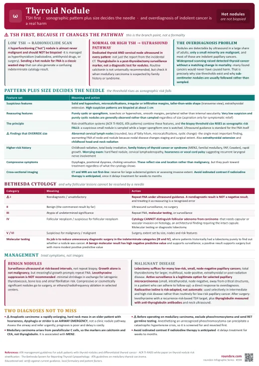
Thyroid nodules are found in a large share of adults who are scanned for something else, and the overwhelming majority are benign. The job is not to biopsy everything, it is to combine sonographic pattern with size to decide who needs a needle, and to check a TSH first because a hyperfunctioning nodule takes a completely different path. Overdiagnosis of indolent papillary cancer is a real harm, not a hypothetical one.
🔍 Overview
The Scale of the Problem
- Nodules are extremely common and mostly incidental -palpable in a minority of adults but detectable by ultrasound in a large share, rising with age and more common in women. Most are found on imaging done for another reason ("incidentalomas").
- Only a small minority are malignant, and of those the great majority are papillary thyroid carcinoma, which is typically indolent with excellent long-term survival.
- Overdiagnosis is the central tension. Widespread scanning has driven a large rise in detected thyroid cancer without a comparable change in mortality -meaning many detected cancers would never have caused harm. That is why guidelines set size thresholds and discourage reflexive biopsy of small nodules.
History and Exam Features That Matter
- Higher-risk history: childhood head and neck radiation, total body irradiation, family history of thyroid cancer or a syndrome (MEN2, familial medullary thyroid cancer, FAP, Cowden), and rapid growth.
- Worrying exam findings: a hard, fixed nodule, cervical lymphadenopathy (the single most concerning exam finding), hoarseness or vocal cord palsy suggesting recurrent laryngeal nerve involvement, and obstructive symptoms.
- Compressive symptoms -dysphagia, positional dyspnea, choking sensation- reflect size and location rather than malignancy, but they change management toward treatment regardless of cytology.
- Symptoms of thyroid dysfunction point toward a functioning nodule or coexisting thyroid disease and change the first test.
🧪 Workup & Imaging
Order TSH first, before anything else, because a low TSH changes the entire pathway. If TSH is suppressed, get a radionuclide thyroid scan: a hyperfunctioning ("hot") nodule is almost never malignant and should NOT be biopsied -it is treated as hyperthyroidism (radioiodine, antithyroid drugs or surgery). Sending a hot nodule for fine-needle aspiration is a classic wasted, and occasionally misleading, step. If TSH is normal or high, proceed down the ultrasound pathway.
Initial Tests
- TSH -the only routine blood test. Thyroglobulin is not a diagnostic test for nodules (it is a post-thyroidectomy cancer surveillance marker), and routine calcitonin measurement is not universally recommended, though it is checked when medullary carcinoma is suspected by family history or syndrome.
- Dedicated thyroid and cervical lymph node ultrasound in every patient with a nodule -it characterizes the nodule, measures it, and surveys the neck nodes, which independently changes management if abnormal.
- CT and MRI are not first-line and are reserved for large substernal goiters or assessing extent of invasive disease. Avoid iodinated contrast if radioiodine therapy is anticipated, as it delays treatment for weeks to months.
Sonographic Risk Features
- Suspicious: solid and hypoechoic, microcalcifications, irregular or infiltrative margins, taller-than-wide shape (on transverse view), extrathyroidal extension, and abnormal cervical nodes.
- Reassuring: purely cystic or spongiform appearance, isoechoic or hyperechoic, smooth margins, peripheral rather than internal vascularity.
- Risk-stratification systems (ACR TI-RADS and the ATA sonographic patterns) combine these features into a risk category, and the biopsy threshold size rises as the sonographic risk falls -a highly suspicious small nodule is sampled while a larger spongiform one is watched.
- Purely cystic nodules do not need FNA for cancer purposes; aspiration is done for symptomatic relief.
Deciding on Fine-Needle Aspiration
- The principle: combine sonographic pattern with maximum diameter. High-suspicion patterns are biopsied at about 1 cm; intermediate patterns at slightly larger sizes; low-suspicion patterns at larger sizes still; and very low suspicion or purely cystic nodules are generally observed rather than sampled regardless of size (unless symptomatic).
- Biopsy regardless of size thresholds when there are abnormal cervical lymph nodes, extrathyroidal extension, or a high-risk history such as childhood radiation exposure -these override the usual size rules.
- Ultrasound guidance is standard, improving adequacy and accuracy over palpation-guided sampling.
- Sub-centimeter nodules are usually not biopsied even with suspicious features, precisely because of the overdiagnosis problem -they are followed instead unless nodes are abnormal or there is extrathyroidal extension.
🔬 Cytology
The Bethesda System
| Category | Meaning | Approximate malignancy risk | Usual action |
|---|---|---|---|
| I | Nondiagnostic / unsatisfactory | Not interpretable | Repeat FNA with ultrasound guidance -a nondiagnostic result is not a negative result, and treating it as reassuring is a recognized error. |
| II | Benign | Very low (a few percent) | Ultrasound surveillance; no surgery. The most common result by far. |
| III | Atypia of undetermined significance (AUS/FLUS) | Low to intermediate | Repeat FNA, molecular testing, or surveillance; molecular markers help avoid diagnostic surgery. |
| IV | Follicular neoplasm / suspicious for follicular neoplasm | Intermediate | Cytology cannot distinguish follicular adenoma from carcinoma -that requires capsular or vascular invasion on histology. Molecular testing or diagnostic lobectomy. |
| V | Suspicious for malignancy | High | Surgery (lobectomy or thyroidectomy depending on features). |
| VI | Malignant | Very high | Surgery, with extent determined by size, nodes and risk features. |
Molecular Testing
- Its job is to reduce unnecessary diagnostic surgery in the indeterminate categories (Bethesda III and IV), where historically many patients had a lobectomy purely to find out whether a nodule was cancer.
- A negative (benign) molecular result has a high negative predictive value and supports surveillance instead of surgery; a positive result supports proceeding to surgery, though its positive predictive value is more modest.
- It does not replace clinical judgment -sonographic appearance, size, growth, symptoms and patient preference all still count.
🚨 Management
Benign Nodules
- Surveillance ultrasound rather than repeat biopsy, with the interval based on sonographic risk -higher-suspicion appearances are followed more closely, and low-risk nodules can be followed at long intervals or eventually stopped.
- Growth alone is not malignancy, but meaningful growth (conventionally an increase of at least 20% in two dimensions with a minimum absolute increase, or a substantial volume change) prompts repeat FNA.
- Levothyroxine suppression therapy is not recommended for benign nodules in iodine-sufficient populations -minimal shrinkage at the cost of iatrogenic thyrotoxicosis, with its bone and atrial fibrillation risks.
- Treat symptoms, not images: compressive or cosmetically significant nodules can be managed with surgery, and in selected centers with ethanol ablation for cystic lesions or radiofrequency ablation for solid benign nodules.
Malignant and Indeterminate Disease
- Surgery is the primary treatment -lobectomy is appropriate for many low-risk, small, node-negative papillary cancers, with total thyroidectomy reserved for larger tumors, multifocal or bilateral disease, nodal involvement, extrathyroidal extension, or prior radiation.
- Active surveillance is a legitimate option for selected papillary microcarcinomas (small, intrathyroidal, node-negative, not adjacent to critical structures) in patients who can adhere to follow-up -a direct response to the overdiagnosis problem.
- Radioactive iodine is risk-adapted, not automatic: it is used selectively in intermediate and high-risk disease rather than routinely for low-risk papillary cancer.
- After thyroidectomy: levothyroxine replacement with a TSH target set by recurrence risk, and surveillance using thyroglobulin with anti-thyroglobulin antibodies plus neck ultrasound.
Two Diagnoses Not to Miss
- Anaplastic thyroid carcinoma -a rapidly enlarging, hard neck mass in an older patient, often with hoarseness, dysphagia or stridor. This is an emergency with a very poor prognosis; airway assessment and urgent referral come before any leisurely nodule pathway.
- Medullary thyroid carcinoma -arises from parafollicular C cells, so calcitonin and CEA are the markers, not thyroglobulin. It is associated with MEN2, so a confirmed diagnosis requires RET germline testing and screening for pheochromocytoma before any surgery -operating on an unrecognized pheochromocytoma is a catastrophic error, which links this topic to the pheochromocytoma page.
🏥 Rounds
Pimp Questions
❓ Why is TSH the first test in a patient with a thyroid nodule, and what changes if it is low?
Because a suppressed TSH sends the patient down a completely different pathway. Low TSH prompts a radionuclide scan, and a hyperfunctioning ("hot") nodule is almost never malignant and should NOT be biopsied -it is managed as hyperthyroidism with radioiodine, antithyroid drugs or surgery. Only if TSH is normal or elevated does the ultrasound-and-FNA pathway apply. Sending a hot nodule for fine-needle aspiration is a classic wasted step that can also generate a confusing indeterminate cytology result.
❓ Why can cytology diagnose papillary carcinoma but not follicular carcinoma?
Because the diagnoses rest on different evidence. Papillary carcinoma is diagnosed by nuclear features -nuclear grooves, pseudoinclusions, chromatin clearing- which are visible in aspirated cells. Follicular carcinoma is distinguished from follicular adenoma only by capsular or vascular invasion, which is an architectural finding requiring the intact capsule on histology, and an aspirate cannot show it. That is why Bethesda IV ("follicular neoplasm") historically meant diagnostic lobectomy, and why molecular testing exists mainly to spare patients that operation.
❓ A 1.2 cm nodule looks spongiform; another patient has a 1.1 cm solid hypoechoic nodule with microcalcifications and an irregular margin. Do both get biopsied?
No -the sonographic pattern sets the size threshold. The solid hypoechoic nodule with microcalcifications and irregular margins is high-suspicion and is biopsied at about 1 cm. The spongiform nodule is a very low-suspicion pattern and is generally observed rather than sampled at that size. The underlying principle is that the biopsy threshold rises as sonographic risk falls, which is how guidelines limit the overdiagnosis of indolent cancers while still catching the ones that matter.
❓ What single exam or ultrasound finding most changes management regardless of nodule size?
Abnormal cervical lymph nodes. Suspicious nodes -rounded shape, loss of the fatty hilum, microcalcifications, cystic change or abnormal vascularity- override the usual size thresholds and warrant FNA of both the node and the nodule, because nodal disease changes staging and the extent of surgery. This is why every thyroid ultrasound should include a survey of the cervical nodes rather than imaging the nodule alone. Extrathyroidal extension and a high-risk history such as childhood radiation similarly override size rules.
❓ Before operating on medullary thyroid carcinoma, what must be excluded and why?
Pheochromocytoma, because medullary thyroid carcinoma is a feature of MEN2, where the two coexist. Inducing anesthesia in a patient with an unrecognized pheochromocytoma can precipitate a catastrophic hypertensive crisis, so biochemical screening comes first, and the pheochromocytoma is resected before the thyroid. A confirmed medullary carcinoma also requires RET germline testing, with family screening implications. Note the marker difference too: calcitonin and CEA, not thyroglobulin, since the tumor arises from parafollicular C cells.
Sample Presentation
Ms. Adeyemi is a 47-year-old woman whose 1.4 cm right thyroid nodule was found incidentally on a CT done after a car accident. She has no compressive symptoms, no hoarseness, no radiation history and no family history of thyroid disease. TSH is 1.8 (normal). Dedicated thyroid ultrasound shows a solid hypoechoic nodule with microcalcifications and an irregular margin, and no abnormal cervical lymph nodes.
Key Points: (1) The normal TSH means no radionuclide scan is needed and the ultrasound-and-FNA pathway applies -had the TSH been suppressed, a hot nodule would have been treated as hyperthyroidism rather than biopsied. (2) This is a high-suspicion sonographic pattern (solid, hypoechoic, microcalcifications, irregular margins), and at 1.4 cm it clears the roughly 1 cm threshold, so ultrasound-guided FNA is indicated. The normal nodes are reassuring but would have overridden size thresholds if abnormal. (3) Counsel her honestly before the result: most nodules are benign, and even if this is papillary carcinoma, many are indolent -lobectomy suffices for many low-risk tumors, and active surveillance is an option for selected microcarcinomas.
Clinic Checklist
- TSH checked first -suppressed TSH means radionuclide scan, and a hot nodule is not biopsied.
- Dedicated thyroid AND cervical node ultrasound obtained, not just a report from the incidental scan.
- Sonographic pattern documented (solid/hypoechoic, microcalcifications, margins, shape) and matched to the size threshold.
- Override features sought: abnormal nodes, extrathyroidal extension, childhood radiation, family syndrome.
- Nondiagnostic cytology repeated, not treated as benign.
- Indeterminate cytology considered for molecular testing before defaulting to diagnostic surgery.
- Levothyroxine suppression not prescribed for benign nodules.
- Rapidly enlarging hard mass with hoarseness or stridor treated as urgent -anaplastic carcinoma is an airway problem, not a clinic problem.
📋 Summary
Summary
Most Nodules Are Benign, and Overdiagnosis Is Real
Nodules are detectable by ultrasound in a large share of adults and only a small minority are malignant, most of those indolent papillary cancers. Widespread scanning raised detected thyroid cancer without a matching mortality change, meaning many found cancers would never have caused harm -which is exactly why size thresholds exist and why reflexive biopsy of small nodules is discouraged.
TSH First, Always
A suppressed TSH sends the patient down a different pathway entirely: get a radionuclide scan, and a hyperfunctioning "hot" nodule is almost never malignant and should NOT be biopsied -it is treated as hyperthyroidism. Only with a normal or elevated TSH does the ultrasound-and-FNA pathway apply. Thyroglobulin is a post-thyroidectomy surveillance marker, not a diagnostic test for nodules.
Pattern Plus Size Decides the Needle
Suspicious: solid and hypoechoic, microcalcifications, irregular or infiltrative margins, taller-than-wide, extrathyroidal extension. Reassuring: purely cystic or spongiform, smooth margins. The biopsy threshold size rises as sonographic risk falls -high-suspicion patterns are sampled around 1 cm while very low-suspicion and purely cystic nodules are observed.
Some Findings Override Size
Abnormal cervical lymph nodes are the single most important finding and warrant FNA of node and nodule regardless of size, because nodal disease changes staging and surgical extent -which is why every thyroid ultrasound must survey the neck. Extrathyroidal extension and childhood head-and-neck radiation similarly override the usual thresholds.
Read Bethesda Correctly
Nondiagnostic (I) is not benign -repeat it under ultrasound guidance, because treating it as reassuring is a recognized error. Benign (II) is the commonest result and goes to surveillance. III and IV are the indeterminate middle, where molecular testing earns its place. V and VI go to surgery.
Why Follicular Lesions Are Different
Papillary carcinoma is diagnosed on nuclear features visible in an aspirate; follicular carcinoma is distinguished from adenoma only by capsular or vascular invasion, an architectural finding that requires the intact capsule on histology. Cytology therefore cannot resolve it, which is why Bethesda IV historically meant diagnostic lobectomy -and why molecular testing exists mainly to spare that operation.
Treat Symptoms, Not Images
Benign nodules get surveillance ultrasound at risk-based intervals, with repeat FNA for meaningful growth (growth alone is not malignancy). Levothyroxine suppression is not recommended -minimal shrinkage in exchange for iatrogenic thyrotoxicosis, bone loss and atrial fibrillation risk. Compressive or cosmetically significant nodules are treated with surgery, or ablation in selected centers.
Two Diagnoses Not to Miss
Anaplastic carcinoma: a rapidly enlarging hard mass in an older patient with hoarseness, dysphagia or stridor is an airway emergency, not a clinic pathway. Medullary carcinoma: markers are calcitonin and CEA, not thyroglobulin, and because of the MEN2 association it requires RET testing and exclusion of pheochromocytoma BEFORE surgery -anesthetizing an unrecognized pheochromocytoma is catastrophic.